Hernia Site Trocar, Case Report and Review.
- 1. Enrique David Gómez Tagle Morales
- 2. Residence of Advanced Laparoscopy, Hospital Angeles del Pedregal, México
Abstract
The trocar site hernia has an incidence of 1.5 to 1.8%. Dependent causes of the surgical technique are: facial length greater than 10 milimeters incision, leaving open the facial defect and its elongation due to prolonged surgical manipulation. 86.3% of cases appears in holes equal to or greater than 10 milimeters. The manipulation of facial defect in procedures lasting more than 80 minutes may increase the risk of a hernia. Early cases of trocar site hernia occuronaverange within 5 days after Surgery manifesting itself in 76.2% with symptoms of intestinal obstruction and presenting as a Richter´s hernia in 46.7%. Suspecting the diagnosis is documented by a CT scan. The most commonly used approach for correction is the facial closing.
Objective and method: To present a case report and transmitupdated information obtained throught review of available literatura to May 2016.
Case report: Female, 68 years. In this third day of post operative period of laparoscopic appendectomy for complicated apendicitis that requiered placement of a penrose externalizad through the left port, ir presents intestinal occlusion, in reintervention trocar site Richter´s hernia was reduced bylaparoscopy closing the facial defect.
Conclusions: The trocar site hernia can be prevented by careful and complete closure of facial defects and using blunttrocarss. The trocar site hernia can give early postoperative intestinal obstruction requiring immediate intervention.
Keywords
• Hernia
• Trocar
Citation
Cortés LA, Tagle Morales EDG (2016) Hernia Site Trocar, Case Report and Review. JSM Gen Surg Cases Images 1(3): 1014.
ABBREVIATIONS
CT: Computerized Tomography; MM: Millimeters; HR: Hour
INTRODUCTION
Mario and Ruchman in 1991 released the first report of trocar site herniation with small bowel obstruction [1]. Swank [2] and Helgstrand [3] found an incidence of hernias trocar site from 0 to 5.2%, while for Mayol [4] and Nassar [5]was 1.5 to 1.8%. Pereira found that the average age for adults with trocar site herniation is 63 years [6]. Montz [7] concluded a direct relationship between the size of the incision and the risk of subsequent herniation based on the 86.3% appear in greater than or equal incisions to 10 millimeters, 10.9% at least 8 mm and 2.7% in less than 8 mm. Kadar [8] found that the closure of minor facial defects of 12 mm significantly decreases the rate of development of a trocar site hernia. The highest incidence of hernias trocar at umbilical level (75.7%) is related to that most 10-mm trocars are placed in the umbilical region [7]. Facial weakness in this region occurs in 12% of people [5,9]. Nassar (5) observed that the elongation and forced dilation of facial incisions of 5 mm, as well as excessive manipulation in a longer operating time of 80 minutes [6] increase the risk of hernia. Duron [10] postulate that with removal trocars a partial vacuum is created in the channel of the trocar favoring the insertion of omentum or intestine with possibility of entrapment. Suggests opening the valves of the trocars to allow progressive removal of CO2. The trocar site hernia early onset usually occurs in the first 5 days after surgery and frequently manifests itself as partial bowel obstruction ( 76.5% ) as a hernia Richter [11] as discovered Sanz and Lopez, with clinical manifestation of vomiting or nausea, bloating and abdominal pain [12] Figure (1).
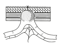
Figure 1 Hernia early-onset type Richter. Taken from Tonouchi H. Ohmori Y. Kobayashi M. Trocar site herniation. Ann Sur 2004.
Hernia early-onset type Richter. The diagnosis is documented by CT in most cases, but can also be performed gastrointestinal contrast study [11]. Pereira indicated that simple or laparoscopic manual reduction with face seal (84%) compared to laparotomy (16%) is most commonly used for correction trocar site hernia [6]. The closure of the facia including the peritoneum in the larger incisions same or greater than 10 mm [3] and ports under 8 mm with long surgical manipulation time are the most important to prevent trocar site herniation measures [13]. It documented that the use of blunt trocars significantly reduces the appearance of hernias trocar in 1.83 to 0.17% [14].
CASE PRESENTATION
It is female patient 68 years old who was admitted with abdominal pain 5 days of evolution; given the characteristics of the picture and the persistence of symptoms it is valued and diagnosed with complicated appendicitis. Intraoperative findings support the diagnosis of perforated appendicitis with free fluid in the cavity of about 400cc of purulent fluid, laparoscopic appendectomy is performed and placement of Penrose drain 1/2 in left port without other eventualities. It presents ileus so the start is performed orally at 48 hr after surgery. At 72 hr starts with oral intolerance, vomiting, intestinal characteristics, accompanied by bloating and cramping abdominal pain, so abdominal scan is done showing intestinal occlusion wall hernia site Penrose drain. Laparoscopy is performed by finding hernia site trocar lateral Richter-type handle partially contained small intestine without gangrene, the hernia is reduced and the facial defect is closed leaving simple points skin, presenting a good evolution after re-intervention Figures (2-5).
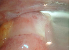
Figure 2 Hernia site trocar in situ.
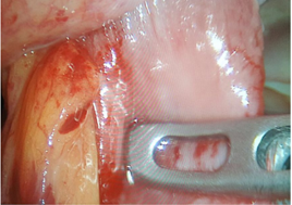
Figure 3 Reduction of intestinal loop.
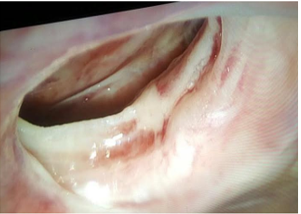
Figure 4 Peritoneal and aponeurotic defect.
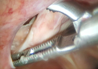
Figure 5 Measurement of aponeurotic defect.
DISCUSSION
In our clinical case causes herniated trocar site are: length greater than 10 millimeters facial incision, lack of facial hole end (to externalize the Penrose) and elongation prolonged handling. In this case an intestinal occlusion occurs in the third postoperative day, which classifies as early onset hernia Richter type. The way to prevent hernia site trocar in this patient could have been: close the facial defect greater than 8 mm, externalize the Penrose by a separate small hole, using a blunt trocar gradually make emptying the pneumoperitoneum. However the technical impact of the trocar site herniation is so small that the safety and feasibility of these measures open opportunities to be supported in clinical studies with appropriate designs and statistical analysis.
ACKNOWLEDGEMENTS
Espacio para agradecimientos.


