Paracolic Collection from Gall Bladder Perforation Ending into a Fatal Liver Complication.
- 1. Colorectal Registrar Sandwell Hospital, UK
Citation
Eltair M (2017) Paracolic Collection from Gall Bladder Perforation Ending into a Fatal Liver Complication. JSM Gen Surg Cases Images 2(4): 1037.
INTRODUCTION
Gall Bladder perforation is life threatening event associated with increased morbidity and mortality. Cholecystectomy with peritoneal lavage is the treatment of choice [1]. A study shows that gallbladder perforation accounts for 3–15% in acute calculus cholecystitis. The commonest site of perforation is fundus of the gallbladder [2]. Even though laparoscopic cholecystectomy (LC) has become the customary method for treating gallstones, some incidents and complications appear rather more frequently than with the open technique. Several aspects of these complications and their treatment possibilities are analysed [3].
CASE PRESENTATION
A 77 years old gentleman presented to the surgical assessment unit complaining of two days shifting pain from epigastrium to umbilicus to right iliac fossa worse on movement while open bowel. The patient was known to have hypertension, type 2 diabetes mellitus and was previously admitted with cholecystitis. On examination, there was decreased air entry on the chest bilaterally; the abdomen was distended, tender on right iliac fossa but soft. The bloods showed WCC 15.5 109 /L, CRP 95 mg/L, ALP 148 U/L, ALT 209 U/L, bilirubin 10 μmol/L, amylase 42 U/L. CXR showed left basal consolidation of the lung with prominent right hilum and normal AXR. The patient was treated as possible appendicitis, given antibiotics, scheduled for CT abdomen and pelvis after being consultedwith suspicion of diverticulitis. The CT was done on the same day of admission showing large collection of right flank around caecum and ascending colon but the appendix was not seen (Figure 1 & 2),
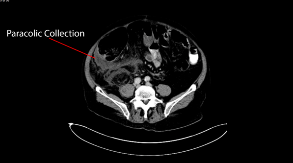
Figure 1 The admission CT showing the paracolic collection.
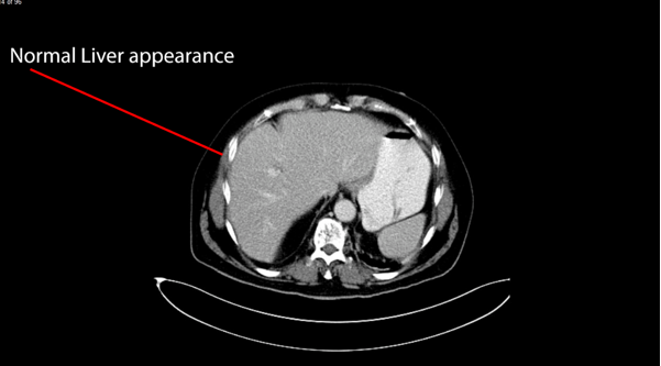
Figure 2 The same day CT showing normal liver.
contracted gall bladder containing stone with common bile duct dilatation, 1.4 cm incidental left adrenal adenoma. The patient was taken on the same night by the consultant on call for laparotomy based on the CT findings with differential diagnosis of perforated appendicitis / perforated cholecystitis. The operative findings were distended large bowel with small amount of bile in the abdomen, the right paracolic gutter and retroperitoneal space were stained with bile so the right colon was mobilized to find the source of bile and the gall bladder was queried for being the source. An Upper GIT consultant was summoned as the primary consultant did not find proper planes and was wondering whether the situation was of colonic perforation or a gall bladder fistulation. The upper GIT surgeon thought the gall bladder was perforated so he did a subtotal cholecystectomy, oversewing of gall bladder remains, washout, leaving two drains in the subhepatic and the paracolic space. The patient went to a ward postoperatively then the next day developed oliguria with elevated liver function tests ALT being 2844 U/L and ALP 307 U/L. The following day, he dropped his Haemoglobin to 69 g/L thus taken by intensive care unit and a CT abdomen and pelvis was done (Figure 3)
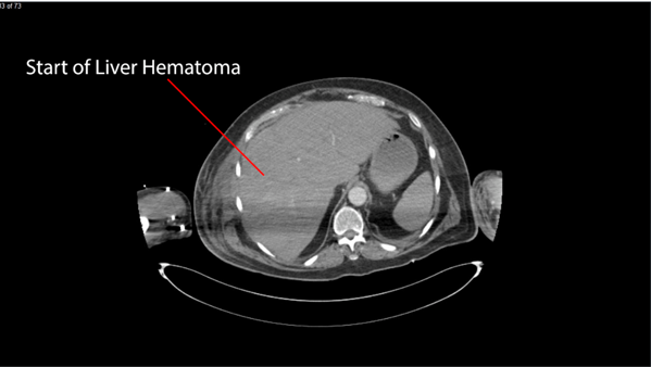
Figure 3 The post operative CT suggesting beginning of liver hematoma.
showing active bleeding in the right lobe of the liver from a relatively distal branch of the right portal vein with intraparenchymal liver haematoma present, possible bile/haematoma collection presence in the right lobe and the right paracolic gutter. The patient remained in ITU for a week without improvement then started desaturating and required intubation so had a CT head which was unremarkable and CT abdomen (Figure 4)
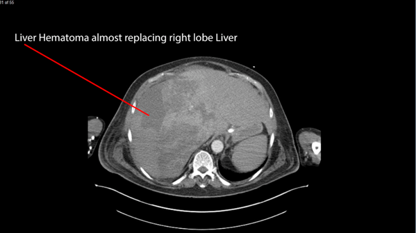
Figure 4 The last CT showing the hematoma almost replacing the right lobe.
which showed increase in the liver subcapsular hematoma with very large liver hematoma in right lobe. The tertiary centre for liver surgery unit was alerted and advised to continue with conservative treatment. The patient remained in ITU with further deterioration for a further week with a drop of Haemoglobin to 68 which required 5 units of blood and 2 units of FFP to keep patient stable. Additional CT abdomen and pelvis in the consecutive days showed deterioration of the liver hematoma and thus the patient was accepted and transferred to the tertiary liver unit.
Table 1: Abbreviations and normal levels glossary.
| Abbreviations | Normal levels |
| WCC: white cell count | <1 mg/L |
| CRP: C-reactive protein | <1 mg/L |
| ALP: alkaline phosphatase | 20-130 U/L |
| ALT: alanine transferase | <41 U/L |
| LC: laparoscopic cholecystectomy CXR: chest X-ray AXR: abdominal X-ray CT: Computerized tomography GIT: gastrointestinal tract ITU: intensive therapy unit FFP: Fresh Frozen Plasma |
bilirubin(normal <21 μmol/L ) amylase(normal <110 U/L ) Haemoglobin (normal 120-180 g/L) |
THE LEARNING POINTS
1. Suspected clinical diagnosis of appendicitis can be masquerading more serious problems.
2. Would there be something else that could have been done in the management whether operative or postoperative in a district hospital
3. What was the cause of the liver pathology postoperatively? Was it a direct Liver injury or tying of a hepatic artery with subsequent right hepatic lobe necrosis?
4. Would the patient have benefited from an early transfer to a liver unit in a tertiary hospital?


