Reversal of Fetal Autoimmune Atrioventricular Block in an Anti-SSA/Ro-Positive Pregnancy Following Combined Immunomodulatory Therapy: A Case Report
- 1. Department of Rheumatology and Immunology, The First Affiliated Hospital of Soochow University, China
- #. Both the authors contributed equally
Abstract
Background: Fetal autoimmune atrioventricular block (AVB) is a rare but severe manifestation of cardiac neonatal lupus, primarily caused by maternal anti-SSA/Ro and anti-SSB/La antibodies crossing the placenta. Second-degree type II AVB often progresses rapidly to complete AVB with high mortality, and few cases demonstrate reversal.
Case Presentation: We report a 33-year-old Asian woman with systemic lupus erythematosus (SLE) and positive anti-SSA/Ro and anti-SSB/La antibodies whose fetus developed second-degree type II AVB at 23 weeks + 3 days gestation. Combined therapy with hydroxychloroquine, dexamethasone, intravenous immunoglobulin (IVIG), and heparin, along with calcium and vitamin D supplementation, resulted in restoration of normal fetal atrioventricular conduction. Serial fetal echocardiography and maternal antibody monitoring documented gradual reversal. The treatment course was complicated by borderline oligohydramnios and fetal growth restriction, which were managed with dose adjustments and close monitoring.
Conclusions: This case illustrates that fetal autoimmune AVB may possess a reversible stage if detected early and managed with individualized immunomodulatory therapy. It emphasizes the importance of early diagnosis, serial echocardiographic monitoring, and multidisciplinary management in high risk pregnancies.
Keywords
• Fetal autoimmune atrioventricular block
• Hydroxychloroquine
• Dexamethasone
• Intravenous immunoglobulin
• Maternal systemic lupus erythematosus
• Congenital heart block
• Cardiac neonatal lupus
Citation
Shen X, Chen Q, Wang L, Wu J, Yin Y, et al. (2026) Reversal of Fetal Autoimmune Atrioventricular Block in an Anti-SSA/Ro-Positive Pregnancy Following Combined Immunomodulatory Therapy: A Case Report. J Cardiol Clin Res. 14(2): 1227.
ABBREVIATIONS
AVB: Atrioventricular Block; CHB: Congenital Heart Block; SLE: Systemic Lupus Erythematosus; NLS: Neonatal Lupus Syndrome; HCQ: Hydroxychloroquine; Dex: Dexamethasone; IVIG: Intravenous Immunoglobulin; Mobitz II: Mobitz type II second-degree Atrioventricular Block; FGR: Fetal Growth Restriction; Oligohydramnios: Reduced Amniotic Fluid Volume; ANAs: Antinuclear Antibodies; Anti-dsDNA: Anti-double-stranded DNA antibody; SSA/Ro: Sjögren’s syndrome-related antigen A/Ro; SSB/La: Sjögren’s syndrome-related antigen B/La; Ro52: 52-kDa Ro/SSA autoantigen; TLR: Toll-like receptor; ACR: American College of Rheumatology
BACKGROUND
Fetal autoimmune atrioventricular block (AVB) is one of the most severe manifestations of cardiac neonatal lupus, typically presenting as fetal bradycardia between 12 and 24 weeks of gestation, and is associated with high mortality and long-term complications [1,2]. The risk of congenital heart block (CHB) in fetuses of anti SSA/Ro-positive mothers is approximately 2%, while the recurrence risk rises more than tenfold (15–20%) in women with a history of previously affected fetuses [3-5]. Furthermore, about 15–20% of infants with autoimmune AVB die within the first three years after birth, and most survivors ultimately require permanent pacemaker implantation and long-term cardiac follow-up [2,6].
Current evidence suggests that fetal autoimmune AVB primarily results from maternal autoantibody mediated injury to the fetal cardiac conduction system. Maternal anti-Ro/SSA antibodies, particularly anti-Ro52 antibodies, can bind to antigens exposed on the surface of apoptotic fetal cardiomyocytes, leading to macrophage activation, inflammatory cytokine release, and profibrotic signaling. If this inflammatory process persists, it may result in irreversible fibrosis, scarring, and calcification of the atrioventricular node, thereby causing permanent conduction abnormalities [7-10]. Therefore, immune mediated AVB may have a limited therapeutic window during the early inflammatory stage, before established fibrosis occurs.
Since first- or second-degree immune-mediated fetal AVB can rapidly progress to complete AVB with poor prognosis [11], early recognition and intervention are critical. However, the optimal management strategy for fetal autoimmune AVB remains controversial. The 2020 American College of Rheumatology (ACR) guidelines recommend sequential fetal echocardiographic monitoring from 16–18 to 26 weeks of gestation and emphasize the continued use of hydroxychloroquine (HCQ) during pregnancy in the absence of contraindications [12]. HCQ is generally considered a preventive therapy, particularly in anti-SSA/Ro-positive pregnancies and in women with a previously affected fetus. Fluorinated corticosteroids, such as dexamethasone, can exert anti-inflammatory effects via the placenta and theoretically suppress fetal cardiac inflammation, and may be considered in selected cases of first- or second-degree AVB. However, robust clinical evidence supporting their ability to reverse or prevent AVB progression remains limited, especially once complete AVB has developed [13,14]. Long-term corticosteroid exposure may also increase the risk of adverse pregnancy outcomes, including fetal growth restriction and oligohydramnios [16]. Therefore, once disease activity stabilizes, steroid dosages should be tapered to the lowest effective dose [15]. Adjunctive therapies such as intravenous immunoglobulin (IVIG), plasmapheresis, and anticoagulation have been used in some cases, but evidence supporting their efficacy remains limited [6,16].
Here, we report a case of second-degree Mobitz type II autoimmune fetal AVB in a pregnant woman with systemic lupus erythematosus and anti-SSA/Ro antibody positivity. Fetal atrioventricular conduction returned to normal after combined treatment with HCQ, dexamethasone, IVIG, and adjunctive therapy. However, the treatment course was complicated by fetal growth restriction and oligohydramnios. This case highlights a potential window of reversibility during the inflammatory phase of fetal autoimmune AVB and underscores the need for individualized risk–benefit assessment and intensive fetal surveillance in high-risk pregnancies.
CASE PRESENTATION
A 33-year-old Asian woman, gravida 1 para 0 (G1P0), with a 10-year history of systemic lupus erythematosus (SLE) presented for prenatal care. She tested positive for anti-SSA/Ro and anti-SSB/La antibodies. Her pre pregnancy body mass index (BMI) was 23.34 kg/m² (159 cm, 55 kg). She had a prior history of breast nodule surgery in 2019 and parotid gland resection in 2024 for mucosa associated lymphoid tissue (MALT) lymphoma (Ki-67: 20%). She denied other surgical history, chronic diseases, or relevant family history.
At baseline, SLE was clinically stable under oral prednisone 7.5 mg/day and hydroxychloroquine (HCQ) 0.2–0.3 g/day, with SLEDAI score of 4. Laboratory findings showed positive antinuclear antibody (ANA, 1:1000), strongly positive anti-SSA/Ro52 and anti-SSB/La antibodies, positive anti-dsDNA antibody, weakly positive anti-nucleosome antibody (±), elevated ESR and CRP, and decreased complement levels (C3 0.62 g/L, C4 0.08 g/L). Thyroid function, echocardiography, and chest CT were unremarkable.
The patient conceived naturally. Last menstrual period (LMP) was August 11, 2025 and estimated date of delivery (EDD) was May 18, 2026. Early prenatal screening, including nuchal translucency, serum screening, noninvasive prenatal testing, and amniocentesis for karyotype, were all normal. Complement levels remained low (C3 0.58 g/L, C4 0.07 g/L), and urine protein was minimally elevated (protein/creatinine 0.045). HCQ was continued at 0.3 g/day, along with vitamin D plus calcium supplementation.
At 14WG, the patient’s progesterone levels were within the normal range, and ultrasound confirmed a viable intrauterine pregnancy. Laboratory tests showed elevated D-dimer (0.82 mg/L), low 25-hydroxy vitamin D and complement levels (C3 0.55 g/L, C4 0.08 g/L). She was advised to continue high-risk pregnancy follow-up in the rheumatology clinic, with weekly fetal echocardiography starting at 18WG.
By 18WG, D-dimer further increased to 3.21 mg/L, likely reflecting physiological hypercoagulability, though SLE activity and risks of antiphospholipid syndrome or thrombosis could not be excluded. Low-molecular-weight heparin calcium 5000 AXa IU subcutaneously once daily was initiated until one week before delivery. Follow-up fetal echocardiography showed a viable singleton with normal fetal heart rate of 151 bpm.
At 23 weeks + 3 days of gestation, fetal echocardiography demonstrated sinus bradycardia (atrial and ventricular rates approximately 76 bpm) with a prolonged A-V interval of 409 ms, suggestive of intermittent second degree type II atrioventricular block (AVB) with 2:1 conduction. Mild tricuspid regurgitation was noted, and cardiac structures were otherwise normal (Figure 1). Given the high-risk features—including strongly positive anti-SSA/Ro52 antibodies, history of SLE, and prior parotid MALT lymphoma—a multidisciplinary consultation was conducted. Prednisone 7.5 mg/day was replaced with dexamethasone 8 mg/day, a fluorinated corticosteroid capable of crossing the placenta, and calcium plus vitamin D3 supplementation was continued for bone protection.

Figure 1: Fetal echocardiography at 23+3 weeks of gestation demonstrating sinus bradycardia with second-degree type II atrioventricular block and 2:1 atrioventricular conduction.
After 6 days of dexamethasone therapy, fetal echocardiography showed partial improvement: ventricular rates 71–119 bpm, atrioventricular conduction alternating between 3:2 and 2:1, but predominantly 2:1. Cardiothoracic ratio decreased to 0.33, and cardiac function remained preserved. Given persistent risk of progression to third-degree AVB and poor prognosis if progression occurred, intravenous immunoglobulin (IVIG) 1 g/kg (60 g) was initiated every 3–4 weeks, while dexamethasone was continued and tapered to 4 mg/day after 2 weeks at 8 mg/day. Weekly fetal echocardiography was performed to guide therapy adjustments.
Fortunately, one month after the initial IVIG treatment, the second-degree type II AVB was reversed by 28 weeks + 4 days of gestation. Fetal echocardiography showed an atrial rate of 158 bpm, with most impulses successfully conducted to the ventricles, although occasional dropped beats were observed, approximately corresponding to a 5:4 conduction ratio. The fetal AV conduction showed improvement, now consistent with second-degree type I AVB. Additionally, maternal serum testing demonstrated a reduction in antinuclear antibody (ANA) titer (1:32) and anti-SSA/Ro52 and anti-SSB/La antibody levels (++), which may be associated with IVIG therapy. IVIG treatment was continued at 1 g/kg every 3 weeks, while dexamethasone (DEX) was gradually tapered to 4 mg/day.
At 32WG, after 3 times of IVIG therapy (1g/kg), subsequent weekly echocardiography consistently showed stable sinus conduction (Figure 2). To maintain fetal atrioventricular conduction and support neonatal immune regulation, IVIG was continued every 3 weeks.
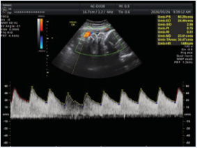
Figure 2: Fetal echocardiography at 32 weeks of gestation demonstrating restoration and maintenance of stable sinus conduction following intravenous immunoglobulin therapy.
During the third trimester, fetal growth slowed, and amniotic fluid index (AFI) gradually decreased to 5.5-7.6 cm. Although the AFI did not meet the diagnostic threshold for overt oligohydramnios, the persistent downward trend indicated borderline oligohydramnios (Figure 3). Given ongoing dexamethasone exposure and concerns about fetal growth restriction and reduced amniotic fluid, dexamethasone was tapered to 2 mg/day. Close fetal monitoring was maintained, including weekly echocardiography, obstetric ultrasound, assessment of amniotic fluid, and evaluation of overall fetal well-being.
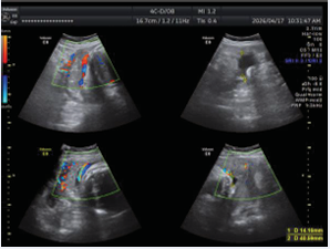
Figure 3: Third-trimester obstetric ultrasound demonstrating borderline oligohydramnios with a progressive reduction in amniotic fluid index.
The longitudinal evolution of fetal AVB and maternal therapeutic interventions are summarized in Figure 4, demonstrating gradual improvement of conduction following IVIG and dexamethasone therapy.
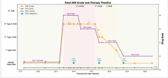
Figure 4: Longitudinal evolution of fetal atrioventricular block and therapeutic interventions throughout gestation.
At 36 weeks of gestation, due to preterm risk and fetal condition, a cesarean section was performed. A low-birth weight infant (2170 g) was delivered with an Apgar score of 10. No neonatal manifestations of lupus were observed. The infant was admitted to the NICU for monitoring, and subsequent cardiology follow-up demonstrated stable cardiac rhythm.
DISCUSSION
Fetal autoimmune atrioventricular block (AVB), particularly second-degree type II, represents one of the most severe cardiac manifestations of neonatal lupus. It is mediated by maternal autoantibodies, primarily anti-SSA/ Ro and anti-SSB/La, crossing the placenta and initiating inflammatory injury in the fetal conduction system. Studies by Buyon et al., demonstrated that once inflammation progresses to fibrosis within the atrioventricular node, reversal becomes exceedingly rare [10,17]. Izmirly et al., reported that most cases of second-degree type II AVB may rapidly progress to complete AVB despite therapy, leading to high mortality and adverse outcomes [2]. Our case highlights a rare instance in which second-degree type II AVB was successfully reversed, emphasizing that early-stage AVB may retain a reversible window prior to irreversible fibrosis.
Fetal immune-mediated AVB is not merely a direct blockade of conduction by maternal autoantibodies, but a multi-step pathological cascade. Maternal anti-SSA/ Ro antibodies bind to apoptotic fetal cardiomyocytes,triggering macrophage activation and downstream inflammatory signaling, including Toll-like receptor (TLR) pathways and type I interferon responses [18,19]. In the early inflammatory phase, edema and immune mediated tissue injury may temporarily impair conduction without causing irreversible structural damage. Persistent inflammation, however, promotes fibrosis and permanent conduction block.
The present case supports this mechanistic framework: serial fetal echocardiography allowed early detection of conduction delay at 23 weeks gestation, and subsequent combined immunomodulatory therapy led to normalization of atrioventricular conduction. This aligns with findings from the PRIDE study (Friedman DM et al.), which suggest that fetuses may remain in a reversible, inflammation-predominant stage if identified early [17]. Continuous fetal echocardiography was crucial for early intervention, reinforcing the importance of dynamic monitoring in high-risk pregnancies.
The 2020 ACR guidelines recommend HCQ for all women positive for anti-SSA/Ro and/or anti-SSB/La antibodies during pregnancy to prevent fetal AVB [12]. In this case, early and continuous HCQ likely played a key disease-modifying role by inhibiting endosomal TLR signaling, particularly TLR7/9 activation, thereby downregulating type I interferon production and limiting inflammatory amplification [9]. Prospective studies by Izmirly et al., demonstrated that HCQ use during pregnancy is associated with significantly reduced recurrence of cardiac neonatal lupus [19].
Fluorinated corticosteroids, especially dexamethasone, bypass placental 11β-hydroxysteroid dehydrogenase type 2 (11β-HSD2) inactivation, cross the placenta, and suppress fetal cardiac inflammation, potentially improving conduction and reducing perinatal adverse outcomes [14]. Their effect primarily involves decreasing inflammatory cytokines and macrophage activation, thereby mitigating edema and immune-mediated damage around the atrioventricular node [20]. However, in cases of third degree complete AVB, fibrosis typically renders reversal impossible, and the clinical efficacy of corticosteroids remains controversial [17]. The ACR guidelines conditionally recommend dexamethasone 4 mg/day for first- or second-degree fetal AVB, while advising against its routine use in isolated third-degree congenital heart block [12]. In our case, dexamethasone treatment improved fetal ventricular rate and facilitated 3:2 conduction, supporting partial reversibility in second-degree type II AVB. Nonetheless, once progression to third-degree AVB occurs, current evidence shows no effective reversal,emphasizing the importance of early intervention. Long-term fluorinated corticosteroid exposure may also increase the risk of placental insufficiency, fetal growth restriction (FGR), oligohydramnios, and potential neurodevelopmental impairment [21]. In our case, borderline oligohydramnios and low birth weight were observed, which could reflect corticosteroid exposure in addition to underlying maternal SLE or antiphospholipid syndrome. Accordingly, dexamethasone was gradually tapered to the lowest effective dose (2 mg/day) once AVB reversal was achieved. Clinical management thus requires dynamic balancing between therapeutic benefits and fetal risk.
IVIG served as adjunctive immunomodulatory therapy, acting through neutralization of pathogenic autoantibodies, Fc receptor modulation, complement inhibition, and downregulation of inflammatory cytokines [22]. Evidence for IVIG in fetal immune-mediated AVB is inconsistent, and standardized protocols are lacking; international guidelines currently recommend IVIG selectively or as supplemental therapy [23]. Initial reports by Kaaja and Julkunen suggested its potential utility [24], and subsequent animal studies, case reports, and small series confirmed IVIG’s effectiveness in preventing progression of immune-mediated fetal AVB [25-29]. However, the PRIDE multicenter prospective trial demonstrated that IVIG at 400 mg/kg every 3 weeks alone did not significantly prevent fetal AVB in high-risk pregnancies [17]. In contrast, in our case, IVIG at 1 g/kg every 3 weeks combined with immunosuppressive therapy successfully reversed second-degree type II AVB and maintained stability, suggesting that optimal IVIG dosing and timing require further investigation.
Heparin was not intended to directly treat fetal AVB but to prevent thrombosis and support placental perfusion, given the propensity for placental microthrombosis in autoimmune pregnancies [30,31]. Calcium and vitamin D supplementation were provided and dynamically monitored to mitigate corticosteroid- and heparin associated maternal bone loss and support fetal development [32].
This case demonstrates that timely recognition of early stage fetal AVB, guided by serial echocardiography, allows for individualized, targeted immunomodulatory therapy. Combining HCQ, corticosteroids, and IVIG may provide an opportunity to reverse conduction abnormalities before fibrosis occurs. Nevertheless, clinicians must balance therapeutic benefits against potential risks, including fetal growth restriction and oligohydramnios associated with corticosteroid exposure, highlighting the need for continuous fetal growth and amniotic fluid monitoring.
As a single-case observational study, the independent contributions of dexamethasone versus IVIG cannot be conclusively determined. Long-term follow-up is limited, and findings may not be generalizable to all fetuses with autoimmune AVB. Additionally, optimal IVIG dosing and timing remain to be clarified in larger, prospective studies.
CONCLUSIONS
Early recognition and proactive combined immunomodulatory therapy—including hydroxychloroquine, dexamethasone, and IVIG—may reverse autoimmune mediated fetal AVB prior to irreversible fibrosis. Careful monitoring, individualized therapy, and multidisciplinary management are essential to optimize maternal-fetal outcomes.
DECLARATIONS
Ethics approval and consent to participate
This study was approved by the Medical Ethics Committee of The First Affiliated Hospital of Soochow University (Approval Number: 2026600). All participants provided written informed consent prior to their inclusion in the study. The research was conducted in accordance with the principles of the Declaration of Helsinki.
Consent for Publication
Written informed consent for publication of this case report and the accompanying images was obtained from the patient. A copy of the written consent form is available for review by the Editor-in-Chief upon request.
Availability of Data and Materials
All data generated or analyzed during this study are included in this published article.
Funding
Application of Suzhou Science and Technology Bureau (No. SKY2023161), Gusu Health Talent Research Project (No. GSWS2023040).
Authors’ contributions
XS and QC contributed equally to this work and are considered co-first authors. XS led the data analysis and drafted the initial manuscript. QC and LW contributed to the data analysis and participated in the preparation of the manuscript. Data collection and patient management were conducted by JW, YY, and TR. JC, TC, and MW conceived and designed the study, supervised the research process, and critically revised the manuscript for important intellectual content. All authors have read and approved the final version of the manuscript for submission.
ACKNOWLEDGEMENTS
We thank the patient and her family for their cooperation and consent to publish this case.
REFERENCES
- Tsuboi H, Sumida T, Noma H, Yamagishi K, Anami A, Fukushima K, et al. Maternal predictive factors for fetal congenital heart block in pregnant mothers positive for anti-SS-a antibodies. Mod Rheumatol. 2016; 26: 569-575.
- Izmirly PM, Saxena A, Kim MY, Wang D, Sahl SK, Llanos C, et al. Maternal and fetal factors associated with mortality and morbidity in a multi-racial/ethnic registry of anti-SSA/ro-associated cardiac neonatal lupus. Circulation. 2011; 124: 1927-1935.
- Buyon JP, Hiebert R, Copel J, Craft J, Friedman D, Katholi M, et al. Autoimmune-associated congenital heart block: Demographics, mortality, morbidity and recurrence rates obtained from a national neonatal lupus registry. J Am Coll Cardiol. 1998; 31: 1658-1666.
- Ambrosi A, Sonesson S-E, Wahren-Herlenius M. Molecular mechanisms of congenital heart block. Exp Cell Res. 2014; 325: 2-9.
- Hansahiranwadee W. Diagnosis and management of fetal autoimmune atrioventricular block. Int J Womens Health. 2020; 12: 633-639.
- Buyon JP, Clancy RM, Friedman DM. Cardiac manifestations of neonatal lupus erythematosus: Guidelines to management, integrating clues from the bench and bedside. Nat Clin Pract Rheumatol. 2009; 5: 139-148.
- Clowse MEB, Eudy AM, Kiernan E, Williams MR, Bermas B, Chakravarty E, et al. The prevention, screening and treatment of congenital heart block from neonatal lupus: A survey of provider practices. Rheumatology (Oxford). Oxford, England; 2018; 57: v9-17.
- Yang X. Clinical features, autoantibodies, and outcome of neonatal lupus erythematosus. Fetal Pediatr Pathol. 2022; 41: 436-442.
- Huang Y, Deng J, Liu J, Yang F, He Y. Autoimmune congenital heart block: A case report and review of the literature related to pathogenesis and pregnancy management. Arthritis Res Ther. 2024; 26: 8.
- Buyon JP, Clancy RM, Friedman DM. Autoimmune associated congenital heart block: Integration of clinical and research clues in the management of the maternal / foetal dyad at risk. J Intern Med. 2009; 265: 653-662.
- Vanoni F, Lava SAG, Fossali EF, Cavalli R, Simonetti GD, Bianchetti MG, et al. Neonatal systemic lupus erythematosus syndrome: A comprehensive review. Clin Rev Allergy Immunol. 2017; 53: 469-476.
- Sammaritano LR, Bermas BL, Chakravarty EE, Chambers C, Clowse MEB, Lockshin MD, et al. 2020 American college of rheumatology guideline for the management of reproductive health in rheumatic and musculoskeletal diseases. Arthritis Care Res (Hoboken). 2020; 72: 461-488.
- Izmirly PM, Saxena A, Sahl SK, Shah U, Friedman DM, Kim MY, et al. Assessment of fluorinated steroids to avert progression and mortality in anti-SSA/ro-associated cardiac injury limited to the fetal conduction system. Ann Rheum Dis. 2016; 75: 1161-1165.
- Sunderji S, Peyvandi S, Jaeggi E, Szwast A, Ryan G, Tessier F, et al. NAFTNet retrospective report on the treatment of anti-ro/SSA mediated fetal heart block with dexamethasone. J Matern Fetal Neonatal Med. 2022; 35: 9263-9270.
- Silver RM, Branch DW. Autoimmune disease in pregnancy. Baillieres Clin Obstet Gynaecol. 1992; 6: 565-600.
- Saxena A, Izmirly PM, Mendez B, Buyon JP, Friedman DM. Prevention and treatment in utero of autoimmune-associated congenital heart block. Cardiol Rev. 2014; 22: 263-267.
- Friedman DM, Kim MY, Copel JA, Llanos C, Davis C, Buyon JP. Prospective evaluation of fetuses with autoimmune-associated congenital heart block followed in the PR Interval and Dexamethasone Evaluation (PRIDE) Study. Am J Cardiol. 2009; 103: 1102-1106.
- Firl CEM, Halushka M, Fraser N, Masson M, Cuneo BF, Saxena A, et al. Contribution of S100A4-expressing fibroblasts to anti-SSA/ro-associated atrioventricular nodal calcification and soluble S100A4 as a biomarker of clinical severity. Front Immunol. 2023; 14: 1114808.
- Izmirly P, Kim M, Friedman DM, Costedoat-Chalumeau N, Clancy R, Copel JA, et al. Hydroxychloroquine to prevent recurrent congenital heart block in fetuses of anti-SSA/ro-positive mothers. J Am Coll Cardiol. 2020; 76: 292-302.
- Popescu MR, Dudu A, Jurcut C, Ciobanu AM, Zagrean AM, Panaitescu AM. A Broader Perspective on Anti-Ro Antibodies and Their Fetal Consequences-A Case Report and Literature Review. Diagnostics (Basel). Basel, Switzerland; 2020; 10: 478.
- Østensen M, Khamashta M, Lockshin M, Parke A, Brucato A, Carp H, et al. Anti-inflammatory and immunosuppressive drugs and reproduction. Arthritis Res Ther. 2006; 8: 209.
- Schwab I, Nimmerjahn F. Intravenous immunoglobulin therapy: How does IgG modulate the immune system? Nat Rev Immunol. 2013; 13: 176-189.
- Liao H, Tang C, Qiao L, Zhou K, Hua Y, Wang C, et al. Prenatal management strategy for immune-associated congenital heart block in fetuses. Front Cardiovasc Med. 2021; 8: 644122.
- Kaaja R, Julkunen H. Prevention of recurrence of congenital heart block with intravenous immunoglobulin and corticosteroid therapy: Comment on the editorial by buyon et al. Arthritis Rheum. 2003; 48: 280-281.
- David AL, Ataullah I, Yates R, Sullivan I, Charles P, Williams D. Congenital fetal heart block: A potential therapeutic role for intravenous immunoglobulin. Obstet Gynecol. 2010; 116: 543-547.
- Ruffatti A, Marson P, Svaluto-Moreolo G, Marozio L, Tibaldi M, Favaro M, et al. A combination therapy protocol of plasmapheresis, intravenous immunoglobulins and betamethasone to treat anti-ro/ la-related congenital atrioventricular block. A case series and review of the literature. Autoimmun Rev. 2013; 12: 768-773.
- Zhao L, Zhou Y, Wang C, Li Y, Zhu Q, Hua Y, et al. Successful prevention of fetal autoimmune-mediated heart block by combined therapies with hydroxychloroquine and intravenous immunoglobulin: A case report. Front Cardiovasc Med. 2021; 8: 759260.
- Yu Y, Du L, Pan J, Zheng J, Chen A, Chen L. A 10-year retrospective study of neonatal lupus erythematous in China. Asian Pac J Allergy Immunol. 2016; 34: 174-178.
- Tran HB, Cavill D, Buyon JP, Gordon TP. Intravenous immunoglobulin and placental transport of anti-ro/la antibodies: Comment on the letter by kaaja and julkunen. Arthritis Rheum. 2004; 50: 337-338.
- Bleau N, Patenaude V, Abenhaim HA. Risk of venous thromboembolic events in pregnant patients with autoimmune diseases: A population-based study. Clin Appl Thromb Hemost. 2016; 22: 285-291.
- Girardi G, Redecha P, Salmon JE. Heparin prevents antiphospholipid antibody-induced fetal loss by inhibiting complement activation. Nat Med. 2004; 10: 1222-1226.
- Andreoli L, Bertsias GK, Agmon-Levin N, Brown S, Cervera R, Costedoat-Chalumeau N, et al. EULAR recommendations for women’s health and the management of family planning, assisted reproduction, pregnancy and menopause in patients with systemic lupus erythematosus and/or antiphospholipid syndrome. Ann Rheum Dis. Elsevier; 2017; 76: 476-485