Acute Kidney Injury in Retroperitoneal Fibrosis: Clinical Characteristics, Management, and Long-Term Renal Outcomes in a Retrospective Cohort
- 1. Department of Nephrology and Internal Medicine, Charles Nicolle Hospital, Tunis Tunisia
- 2. Kidney Pathology Laboratory LR00SP01, Charles Nicolle Hospital, Tunis-Tunisia
- 3. Faculty of Medicine of Tunis, University of Tunis El Manar; Tunis-Tunisia
Abstract
Introduction: Acute kidney injury (AKI) secondary to retroperitoneal fibrosis (RPF) is a rare disorder characterized by excessive retroperitoneal fibrosis leading to urinary tract obstruction. Data specifically addressing patients presenting with severe AKI and their long-term renal outcomes are limited. Moreover, predictors of renal recovery and progression to chronic kidney disease (CKD) or end-stage kidney disease (ESKD) remain poorly defined, partly due to heterogeneity in study populations, therapeutic strategies, and follow-up protocols. We conducted a retrospective study to comprehensively analyze the clinical, biological, radiological, and therapeutic characteristics of this condition and its renal outcomes.
Methods: We retrospectively included 24 patients with AKI associated with RPF. Clinical, laboratory, imaging, histological, and therapeutic data were extracted from medical records for detailed analysis.
Results: The mean age at diagnosis was 50.4 years, with a male predominance. The most frequent presenting symptoms were low back pain (83.3%) and general malaise (79.2%). All patients exhibited renal impairment, with 59% classified as stage 3 AKI. Imaging revealed ureteropyelocal dilatation in all cases, and poor renal differentiation in 23.8%. Histology showed non-specific fibro-inflammatory changes in 27% of cases. Therapeutically, 62.5% required emergency urinary drainage, and 29% required renal replacement therapy. Corticosteroids were administered to 83.3% of patients for a mean duration of 13.8 months. Relapse occurred in 33% during follow-up, and 70.8% progressed to chronic kidney disease.
Conclusion: AKI due to RPF is a rare but severe condition, predominantly affecting middle-aged men. This study provides valuable insights into the clinical course, management, and long-term renal outcomes of patients with severe AKI due to RPF. Early recognition and timely intervention are critical to prevent progression to CKD, and our findings may help identify patients at higher risk of poor renal recovery
Keywords
• Retroperitoneal fibrosis
• Renal function
• Acute Renal Failure
• Renal Prognosis
Citation
Fekih A, Hajji M, Rawnak H, Harzallah A, Barbouch S, et al. (2026) Acute Kidney Injury in Retroperitoneal Fibrosis: Clinical Characteristics, Management, and Long-Term Renal Outcomes in a Retrospective Cohort. J Clin Nephrol Res 13(1): 1128.
ABBREVIATIONS
AAN:Antinuclear Antibodies;AEG: General Condition Deterioration; ANCA: Anti-neutrophil Cytoplasmic Antibodies; CRP: C-reactive Protein; Cs: Corticosteroids; DFGe: Estimated Glomerular Filtration Rate; EER: Extrarenal Replacement Therapy; ECBU: Urine Cytobacteriological Examination; HTA: Arterial Hypertension; FRP: Retroperitoneal Fibrosis; IRA: Acute Kidney Injury; IRC: Chronic Kidney Disease; IRCT: End stage renal Disease; IRM: Magnetic Resonance Imaging; KDIGO: Kidney Disease: Improving Global Outcomes; LES: Systemic Lupus Erythematosus; MDRD: Modification of Diet in Renal Disease; PAS: Systolic Blood Pressure; PAD: Diastolic Blood Pressure; SIB: Biological Inflammatory Syndrome; TDM: Computed Tomography; VS: Erythrocyte Sedimentation Rate
INTRODUCTION
Retroperitoneal fibrosis (RPF) is a rare fibro inflammatory disorder characterized by the development of dense fibrotic tissue in the retroperitoneum, which may entrap adjacent structures, particularly the ureters, leading to obstructive uropathy [1]. The estimated annual incidence ranges from 0.1 to 1.3 cases per 100,000 inhabitants [2].
Approximately two-thirds of cases are considered idiopathic, whereas the remaining cases are secondary to infections, prior abdominal or pelvic surgery, malignancies, certain medications, or inflammatory abdominal aortic aneurysms. In recent years, a significant proportion of previously classified idiopathic cases has been redefined as part of IgG4-related disease, supporting the hypothesis of a systemic immune-mediated pathogenesis in a subset of patients [3].
The clinical presentation of RPF is often insidious and nonspecific, frequently leading to delayed diagnosis. Progressive ureteral encasement may result in obstructive acute kidney injury (AKI), which, if not promptly recognized and managed, can evolve into chronic kidney disease (CKD) or even end-stage kidney disease (ESKD) [4].
Although overall patient survival is generally favorable, renal prognosis remains a major concern. Previous studies have reported CKD rates ranging from 20% to 47% among affected patients [5,6]. However, data specifically addressing patients presenting with severe AKI and their long-term renal outcomes are limited. In addition, predictors of renal recovery and progression to CKD or ESKD remain poorly defined, partly due to heterogeneity in study populations, therapeutic strategies, and follow-up protocols.
We therefore conducted a retrospective cohort study to investigate the clinical presentation, management strategies, and long-term renal prognosis of patients with RPF-associated AKI, and to determine predictors of renal recovery and progression to chronic kidney disease or end-stage kidney disease.
METHODS
Study design and population
We conducted a retrospective descriptive and analytical cohort study in the Department of Internal Medicine A at Charles Nicolle Hospital, Tunis, Tunisia, covering the period from January 1990 to December 2022.
Adult patients (≥18 years) were eligible if they had a diagnosis of retroperitoneal fibrosis (RPF) confirmed by imaging studies and presented with acute kidney injury (AKI) attributable to RPF. All included patients were hospitalized and subsequently followed in our department.
Patients were excluded if they were younger than 18 years, had no radiological evidence of RPF, had a follow-up duration of less than three months, or had incomplete or non-exploitable medical records.
Data collection
Clinical, biological, radiological, therapeutic, and follow-up data were retrospectively extracted from medical records using a standardized data collection form. Variables collected included demographic characteristics, clinical presentation at diagnosis, laboratory findings (serum creatinine, inflammatory markers, hemoglobin level, and calcium–phosphate parameters), imaging features, and histological findings when available.
Therapeutic data included urinary drainage procedures, corticosteroid therapy, immunosuppressive treatment, and renal replacement therapy. Follow-up data comprised renal outcomes, remission status, and relapse events.
Definitions
AKI was defined and staged according to the 2012 Kidney Disease: Improving Global Outcomes (KDIGO) criteria [7], based on any of the following: an increase in serum creatinine ≥0.3 mg/dL (26.5 µmol/L), an increase to ≥1.5 times baseline within seven days, or urine output <0.5 mL/kg/h for at least six hours.
When baseline serum creatinine was unavailable, the acute nature of kidney injury was inferred from preserved renal size and corticomedullary differentiation on imaging, absence of chronic normocytic anemia, and absence of long standing calcium–phosphate abnormalities suggestive of chronic kidney disease.
Renal recovery was defined as a serum creatinine level ≤25% above baseline. In cases with unknown baseline values, recovery was defined as a serum creatinine ≤126 µmol/L. Non-recovery was defined as persistence of serum creatinine >75% above baseline or ongoing need for renal replacement therapy.
Progression to chronic kidney disease (CKD) was defined according to KDIGO recommendations as a persistent reduction in kidney function lasting more than three months [8].
Given the absence of standardized definitions for RPF outcomes, remission was defined as resolution of clinical symptoms, normalization of inflammatory markers, and improvement in renal function. Relapse was defined as recurrence of hydronephrosis and/or a rise in serum creatinine during follow-up.
Statistical analysis
Statistical analysis was performed using SPSS software (version 26.0). Categorical variables were expressed as absolute frequencies and percentages, while continuous variables were presented as mean ± standard deviation or median with range, as appropriate.
Comparisons between categorical variables were performed using Pearson’s chi-square test or Fisher’s exact test, as appropriate. Associations between continuous variables were assessed using Pearson’s correlation coefficient.
Binary logistic regression analysis was conducted to identify independent predictors of progression to CKD. Variables with clinical relevance or statistical significance in univariate analysis were entered into the multivariate model.
A two-sided p-value <0.05 was considered statistically significant. Mortality incidence was calculated as the number of deaths divided by the total follow-up duration.
RESULTS
Between January 1990 and December 2022, 36 patients were followed for retroperitoneal fibrosis (RPF). After application of inclusion and exclusion criteria, 24 patients were retained for analysis.
The median age at diagnosis was 51 years (range 25–78), with 58.3% of patients aged between 40 and 65 years. A marked male predominance was observed (75%), yielding a male-to-female ratio of 3:1.
Hypertension was the most frequent comorbidity (45.8%), and 50% of patients were active smokers. Two patients had a history of malignancy (gastric cancer treated by gastrectomy and untreated prostate cancer), and four had prior abdominal or pelvic surgery.
Clinical presentation
Lumbar pain was the most common presenting symptom, occurring in 20 (83.3%) patients, followed by general deterioration in 19 (79.2%). Hypertension was observed in 15 (62.5%) patients, with a mean systolic blood pressure of 146 ± 29 mmHg and a mean diastolic blood pressure of 81 ± 12 mmHg. Oliguria or anuria occurred in four patients. Proteinuria was detected in six patients and microscopic hematuria in two. Lower limb edema was observed in two cases.
Laboratory findings
Renal impairment at presentation was severe, with a mean serum creatinine level of 650 ± 501 µmol/L and a median estimated creatinine clearance of 9.6 mL/min. Twenty-four-hour proteinuria exceeded 0.5 g in two patients.
Anemia was present in 19 (79.2%) patients and was predominantly inflammatory in origin. Inflammatory markers were elevated in 15 (62.5%) patients. Hypergammaglobulinemia was detected in 66% of tested patients. Immunological screening was largely negative. IgG4 measurement was available in one patient and was within the normal range. Tumor markers were elevated in three patients without confirmed malignant progression.
Imaging and histology
Renal ultrasound revealed bilateral urinary tract dilation in 17 (71.4%) patients. Abdominal computed tomography confirmed uni- or bilateral hydronephrosis associated with a retroperitoneal fibrous mass encasing adjacent vessels and ureters (Figure 1). Magnetic resonance imaging was performed in two patients with contraindications to iodinated contrast.
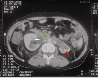
Figure 1: Computed tomography scan showing retroperitoneal fibrosis encasing the retroperitoneal vessels (green arrow), associated with dilation of the excretory cavities more pronounced on the right side (white arrow), and left renal atrophy (red arrow).
Fibrotic tissue biopsy was obtained in nine patients, showing nonspecific fibro-inflammatory changes in eight and retroperitoneal amyloidosis in one.
RPF was classified as idiopathic in 79% of cases, including one patient with IgG4-related disease, and secondary in 20.8% due to malignancy, autoimmune disease, or amyloidosis (Table 1).
Table 1: Etiologies and corresponding medical and surgical management of secondary retroperitoneal fibrosis in the study population
|
Etiology of Secondary RPF |
Number of Patients |
Percentage (%) |
Medical Treatment |
Surgical Treatment |
|
Paraneoplastic |
|
|
|
|
|
Gastric tumor |
1 |
4.1 |
- |
Total gastrectomy |
|
Prostate tumor |
1 |
4.1 |
- |
Patient died before surgical treatment |
|
Autoimmune/inflammatory diseases |
|
|
|
|
|
Systemic lupus erythematosus (SLE) |
1 |
4.1 |
Corticosteroids 1 mg/kg/day for 5 months + Azathioprine for 1 year |
- |
|
Takayasu disease |
1 |
4.1 |
Corticosteroids 1 mg/kg/day for 8 months |
Aortic graft |
|
Amyloidosis |
1 |
4.1 |
Colchicine |
Ureterolysis |
Management
Urgent urinary drainage was required in 15 (62.5%) patients, and 7 (29.2%) required emergency hemodialysis. Corticosteroids were administered in 20 (83.3%) patients at doses ranging from 0.5 to 1 mg/kg/day for a median duration of 8.5 months. Two patients received additional immunosuppressive therapy. Surgical ureterolysis was performed in two cases. The management of secondary RPF was directed toward the underlying cause (Table 1).
Outcomes
The median follow-up duration was 46.5 months. At last follow-up, 7 (29.2%) patients achieved normalization of renal function, whereas 17 (70.8%) progressed to chronic kidney disease (CKD), including 7 (29.2%) who reached stage 5 CKD. The relapse rate was observed in 8 (33.3%) patients. Infectious complications, mainly urinary tract infections related to prolonged stenting, occurred in 6 (25%) patients.
In the analytical study, the overall mortality rate during a cumulative follow-up of 140 patient-years was 2.13% per year. Age and hypertension were independently associated with progression to CKD on multivariate analysis. The annual mortality rate was 2.13% (95% CI: 0.28–4.54).
DISCUSSION
This retrospective cohort study provides a comprehensive evaluation of patients with RPF-associated AKI over a 30-year period. Our findings confirm that RPF predominantly affects middle-aged men and is frequently diagnosed at an advanced stage of renal impairment [9,10].
Clinically, most patients presented with non-specific symptoms, reflecting the insidious onset of RPF. Low back pain was reported in 83.3% of cases, followed by general malaise in 79.2%. These results align with existing literature, which identifies pain as the most frequent presenting symptom and a common reason for delayed diagnosis due to its non-specific nature (9). Renal signs were dominated by hypertension, affecting 62.5% of patients, which may result from extrinsic compression of renal vessels or obstructive uropathy [9].
Approximately 40–50% of patients with retroperitoneal fibrosis present with elevated serum creatinine levels at the time of diagnosis [10]. In our cohort, renal impairment was particularly severe at presentation, with a markedly elevated mean serum creatinine of 650 ± 501 µmol/L and a median creatinine clearance of 9.6 mL/min [2.9–66 mL/ min]. These findings indicate advanced renal dysfunction at diagnosis and likely reflect delayed recognition of the disease, as well as prolonged obstructive uropathy prior to intervention. This likely explains the high rate of CKD progression observed during follow-up.
The majority of cases were classified as idiopathic, consistent with existing literature [3]. IgG4-related disease was identified in only one patient, underscoring the importance of systematic evaluation but also highlighting its relatively low prevalence. Although rare, this association should be actively investigated in patients with RPF, as it may have important implications for both management and therapeutic decisions.
Management relied primarily on prompt urinary drainage combined with corticosteroid therapy. The high rate of emergency dialysis reflects the severity of obstruction-induced AKI at presentation. Immunosuppressive agents are considered therapeutic options in refractory cases or to facilitate corticosteroid-sparing regimens [3]. Surgical intervention, mainly ureterolysis, was required in only two patients, suggesting that most patients can be managed successfully with medical therapy combined with adequate urinary drainage [11,12].
Despite treatment, long-term renal outcomes were suboptimal, with more than two-thirds of patients progressing to CKD. The relapse rate of 33% further emphasizes the need for prolonged monitoring. slightly higher than previously reported (7–50%), likely due to differences in follow-up duration and definitions of recurrence [13,14]. These findings underscore the importance of close clinical and radiologic monitoring, even after apparent remission (Table 2).
Table 2: Comparison of renal outcomes and long-term follow-up across published series of retroperitoneal fibrosis
|
Study |
Country |
Number of Patients |
Mean Follow-up (months) |
Remission (%) |
Relapse (%) |
Chronic Kidney Disease (%) |
|
Moriconi et al. [15] |
Italy |
37 |
90 |
70.2 |
40.5 |
– |
|
Jadhav et al. [16] |
India |
21 |
20.4 |
70 |
17.6 |
– |
|
Laabidi et al. [17] |
Tunisia |
30 |
53.2 |
76 |
53 |
20 |
|
Kermani et al. [18] |
United States |
185 |
48 |
54 |
12 |
32 |
|
Present study |
Tunisia |
24 |
70.3 |
58 |
33 |
70.8 |
Hypertension and smoking were highly prevalent in our cohort. Hypertension may reflect both pre-existing cardiovascular risk and renal vascular compression secondary to fibrosis. Importantly, age and hypertension emerged as independent predictors of CKD progression, in agreement with previously reported risk factors such as baseline creatinine and comorbid conditions [6-17].
Strengths and Limitations
The strengths of this study include the long follow-up period, the relatively large number of well-characterized patients given the rarity of RPF, and the comprehensive clinical and radiological assessment.
However, limitations must be acknowledged. The retrospective, single-center design may introduce selection and information bias. The small sample size limits statistical power and generalizability. Additionally, incomplete immunological and histological evaluation in some patients may have led to underestimation of IgG4 related cases.
Prospective multicenter studies with standardized therapeutic protocols and long-term renal outcome assessment are needed to refine prognostic stratification and optimize management.
DECLARATIONS
Ethics approval and consent to participate
The study was submitted to and approved by the Ethics Committee of Charles Nicolle Hospital, Tunis, Tunisia (Reference number: ETHIQUE-HCN-081-2026).
All procedures performed in this study were in accordance with the ethical standards of the institutional research committee and with the 1964 Helsinki Declaration and its later amendments.
Due to the retrospective nature of the study and the use of anonymized patient data, the requirement for informed consent was waived by the Ethics Committee.
Availability of data and materials
The datasets generated and/or analyzed during the current study are available from the corresponding author on reasonable request.
CONCLUSION
In this retrospective cohort, RPF-associated acute kidney injury was often severe at diagnosis and led to a high rate of progression to chronic kidney disease despite urinary drainage and corticosteroid therapy. Older age and hypertension were independently associated with adverse renal outcomes. These findings underscore the need for early diagnosis and close long-term renal monitoring in patients with RPF.
REFERENCES
- Van Bommel EFH. Retroperitoneal fibrosis. Neth J Med. 2002; 60: 231-242.
- Vaglio A, Maritati F. Idiopathic Retroperitoneal Fibrosis. J Am Soc Nephrol. 2016; 27: 1880-1889.
- Jakhlal N, Elghazoui A, Jabbour Y. Fibrose rétropéritonéale: revue de littérature. Canadian Urological Association J. 2017; 111-112: E26.
- Vaglio A, Salvarani C, Buzio C. Retroperitoneal fibrosis. Lancet. 2006; 367: 241-251.
- ?o? I, Wieliczko M, Lewandowski J. Retroperitoneal Fibrosis Is Still an Underdiagnosed Entity with Poor Prognosis. Kidney Blood Press Res. 2022; 47: 151-162.
- Gallais Sérézal I, Le Jeune S, Belenfant X. Fibrose rétropéritonéale idiopathique : évolution à long terme du pronostic rénal dans une série rétrospective multicentrique de 30 cas. La Revue de Médecine Interne. 2014; 35: 570-576.
- Khwaja A. KDIGO clinical practice guidelines for acute kidney injury. Nephron Clin Pract. 2012; 4: c179-184.
- Stevens PE, Levin A, Kidney Disease: Improving Global Outcomes Chronic Kidney Disease Guideline Development Work Group Members. Evaluation and management of chronic kidney disease: synopsis of the kidney disease: improving global outcomes 2012 clinical practice guideline. Ann Intern Med. 2013; 158: 825-830.
- Gómez RJ, Miguel QL, Mario ÁM. RETROPERITONEAL FIBROSIS: ALITERATURE REVIEW. Archivos Españoles de Urología. 2020; 73: 60-67.
- Raglianti V, Rossi GM, Vaglio A. Idiopathic retroperitoneal fibrosis: an update for nephrologists. Nephrol Dial Transplant. 2021; 36: 1773-1781.
- Chronic periaortitis | Rheumatology | Oxford Academic. 2024.
- Mertens S, Zeegers AGM, Wertheimer PA. Efficacy and complications of urinary drainage procedures in idiopathic retroperitoneal fibrosis complicated by extrinsic ureteral obstruction. Int J Urol. 2014; 21: 283-288.
- Fry AC, Singh S, Gunda SS. Successful use of steroids and ureteric stents in 24 patients with idiopathic retroperitoneal fibrosis: a retrospective study. Nephron Clin Pract. 2008; 108: c213-220.
- Ha YJ, Jung SJ, Lee KH. Retroperitoneal Fibrosis in 27 Korean Patients: Single Center Experience. J Korean Med Sci. 2011; 26: 985-990.
- Moriconi D, Giannese D, Capecchi R. Risk factors for relapse and long-term outcome of idiopathic retroperitoneal fibrosis. Clin Exp Nephrol. 2019; 23: 1147-1153.
- Jadhav KK, Kumar V, Punatar CB. Retroperitoneal fibrosis-clinical presentation and outcome analysis from urological perspective. Investigative and Clinical Urology. 2017; 58: 371.
- JL, Yb A, SC. Retroperitoneal fibrosis: A retrospective review of clinical presentation, treatment and outcomes. Saudi journal of kidney diseases and transplantation : an official publication of the Saudi Center for Organ Transplantation, Saudi Arabia. 2015 ; 21.
- Kermani TA, Crowson CS, Achenbach SJ. Idiopathic retroperitoneal fibrosis: a retrospective review of clinical presentation, treatment, and outcomes. Mayo Clin Proc. 2011; 86: 297-303.