Nephrotic Syndrome Due to Focal Segmental Glomerulosclerosis: Clinical Profile, Histological Variants, and Renal Outcomes
- 1. Internal Medicine Department A, Charles Nicolle Hospital, Tunis, Tunisia
Abstract
Focal segmental glomerulosclerosis (FSGS) is a leading cause of nephrotic syndrome in adults and a major contributor to chronic kidney disease progression. We conducted a retrospective, single-center study including 134 adults with biopsy-proven FSGS and nephrotic syndrome, hospitalized at the Internal Medicine A Department of Charles Nicolle Hospital between 1983 and 2022. The study aimed to describe clinical, histological, and outcome profiles and identify predictors of poor renal prognosis.
The mean age at diagnosis was 35.3 ± 14.6 years, with a male predominance (63.4%). Edema was the most frequent presenting symptom (79.1%), and C3 deposits. 46.3% had renal insufficiency at presentation. Light microscopy revealed flocculo-capsular synechiae in 84.9% of biopsies. Among 28 biopsies classified using the Columbia classification, 17 were Tip lesions and 11 collapsing variants. Immunofluorescence was positive in 87%, most commonly with segmental IgM and
First-line corticosteroid therapy achieved complete remission in 42.5%, partial remission in 23%, and steroid resistance in 34.5%. Relapses occurred in 30.1% of cases. Second-line immunosuppressive therapy was used in resistant or dependent cases. Chronic kidney disease developed in 53.7% of patients, with 33.6% progressing to stage 5 CKD. In multivariate analysis, higher proteinuria at 4 months (OR=1.786) and steroid resistance (OR=3.812) predicted progression to stage 5 CKD, whereas complete remission (OR=0.204) and the tip lesion variant (OR=0.087) were associated with favorable renal outcomes.
Keywords
• Focal segmental
• Glomerulosclerosis
• Nephrotic syndrome
• Renal failure
• Corticosteroid therapy
Citation
Chahed B, Agrebi S, Azzabi A, Sallemi N, Zallema D, et al. (2026) Nephrotic Syndrome Due to Focal Segmental Glomerulosclerosis: Clinical Profile, Histological Variants, and Renal Outcomes. J Clin Nephrol Res 13(1): 1130.
INTRODUCTION
Nephrotic syndrome (NS) is a glomerular syndrome defined by the presence of heavy proteinuria associated with hypoalbuminemia. NS is considered primary when the etiological investigation is negative. In adults, idiopathic NS, which includes two histological entities—minimal change disease (MCD) and primary focal segmental glomerulosclerosis (FSGS) is one of the most common causes of primary NS [1] . FSGS is a histopathological entity characterized by relatively nonspecific glomerular sclerotic and hyaline lesions, distinguished by their segmental and focal distribution. On immunofluorescence, segmental deposits of IgM and C3 may be observed. This histological entity was first described in 1957 [2]. This podocytopathy has shown an increasing prevalence over the past decades [3,4]. Currently, several forms are recognized, including primary, secondary, genetic, and forms of uncertain etiology classified as “undetermined causes” [5]. Primary FSGS often presents with nephrotic syndrome. Its pathophysiology remains incompletely understood. This glomerulopathy is thought to be mainly caused by circulating permeability factors that play a key role in podocyte foot process effacement and are implicated in the risk of recurrence after kidney transplantation. Distinguishing between these different forms is of prognostic importance and is crucial for appropriate management [6].
The Columbia histologic classification, published in 2004, includes five histologic variants: tip lesion, cellular variant, perihilar variant, collapsing variant, and not otherwise specified (NOS). This classification has improved the initial management of FSGS by providing both diagnostic and prognostic information. However, its application may be challenging when interpreting lesions that display mixed features of several variants [7]. Current treatment of primary FSGS relies on the use of immunosuppressive agents, mainly glucocorticoids and calcineurin inhibitors, which act directly on the podocyte phenotype [8,9]. In cases of resistance to immunosuppressive therapy, a genetic form of FSGS should be suspected, as it often presents as steroid-resistant nephrotic syndrome [10].
Despite its significant prevalence, studies focusing on NS associated with FSGS in Tunisia remain limited and include relatively small numbers of patients. Therefore, we conducted a retrospective descriptive study in the Internal Medicine A Department of Charles Nicolle Hospital, including patients hospitalized for the management of NS related to biopsy-proven FSGS.
The primary objective of our study was to analyze the clinical, histological, and outcome profiles of this podocytopathy. The secondary objective was to identify potential factors influencing renal prognosis in order to improve patient management.
METHODS
This was a retrospective, descriptive, single-center study conducted in the Department of Internal Medicine A at Charles Nicolle Hospital, including patients followed for NS with a diagnosis of focal segmental glomerulosclerosis (FSGS) confirmed by renal biopsy over a 39-year period (1983-2022).
The study-included patients hospitalized in Internal Medicine A with NS and histologically confirmed FSGS. Exclusion criteria were: FSGS without nephrotic syndrome at diagnosis, patients initially managed in the pediatric department at the Children’s Hospital, foreign patients, those with another concomitant glomerular disease, patients with follow-up <6 months, or cases with missing medical records.
Collected data included epidemiological characteristics such as age and sex, clinical parameters including edema, blood pressure) as well as laboratory findings including renal function, 24-hour proteinuria, and serum albumin levels. Histopathological data were obtained from renal biopsy analyses performed using light microscopy with standard staining techniques and immunofluorescence. Information on therapeutic interventions was also recorded, including corticosteroid therapy and other immunosuppressive treatments. During follow-up, patients were monitored for treatment response, occurrence of relapses, progression to chronic kidney disease and stage 5 dialysis-dependent CKD, and the need for kidney transplantation.
Statistical Analysis
All data were analyzed using Statistical Package for the Social Sciences (SPSS) version 26. Descriptive statistics were presented as frequencies for categorical variables and as mean, median, standard deviation, and range for continuous variables. Comparative analyses were performed using the Chi-square test or Fisher’s exact test for categorical variables and the Mann-Whitney U test for continuous variables. Multivariate logistic regressIon was conducted to identify independent risk factors for progression to stage 5 D CKD, with adjusted odds ratios (ORs) and 95% confidence intervals (CIs). Statistical significance was set at p < 0.05, and patients were categorized into two groups based on progression to stage 5D (progressors vs. non-progressors).
RESULTS
Between May 1983 and December 2022, nephrotic syndrome secondary to FSGS was diagnosed in 221 patients hospitalized in the Department of Internal Medicine A. Among them, 134 patients were included in the present study.
The mean age at diagnosis of nephropathy was 35.3 ± 14.6 years, ranging from 15 to 63 years, with a median age of 32 years. There was a male predominance, with 63.4% men and 36.6% women, corresponding to a male to-female ratio of 1.73.
The follow-up duration ranged from 6 to 440 months, with a median follow-up of 72 months.
Edematous syndrome was the most common presenting manifestation in our series (79.1%). Nephrotic syndrome was pure in 22 patients (16.4%) and impure in 112 patients (83.6%). Hypertension was observed in 60 patients (44.8%), including five patients with previously known hypertension. Microscopic hematuria was detected in 81 patients (60.4%).
he median 24-hour proteinuria was 5.7 g/day, with values ranging from 3 to 22.9 g/day. Renal insufficiency at presentation was observed in 62 patients (46.3%).
A kidney biopsy was performed in all patients during their hospitalization in the Department of Internal Medicine A. The diagnosis of FSGS was established at the first renal biopsy in 132 patients and at the second biopsy in two patients, who had previously been diagnosed with MCD on the initial biopsy.
Light microscopy findings
The number of glomeruli per biopsy ranged from 5 to 66, with a median of 20 glomeruli. The percentage of globally sclerotic glomeruli per biopsy ranged from 0% to 75%, with a median of 5.5%.
The most frequent histological lesion was the presence of glomerulotubular adhesions (flocculo-capsular synechiae), observed in 84.9% of renal biopsies (Figure 1). Isolated glomerular involvement was identified in 36 patients (26.9%).

Figure 1: Flocculo-capsular synechia (Light microscopy ×400, Silver stain) in a 16-year-old patient hospitalized for evaluation of an atypical nephrotic syndrome
Tubulointerstitial involvement was observed in 93 cases (69.4%), including interstitial fibrosis in 61.9% and tubular atrophy in 38.8% of cases. Vascular lesions were found in 61 patients (45.5%).
The Columbia histopathological classification was applied in 28 renal biopsies, revealing 17 cases of the tip lesion variant and 11 cases of the collapsing variant (Figure 2 and Figure 3).

Figure 2: Tip lesion–type focal segmental glomerulosclerosis (Light microscopy ×400, Masson’s trichrome) in a 23-year-old patient hospitalized for evaluation of an atypical nephrotic syndrome
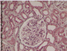
Figure 3: Collapsing-type focal segmental glomerulosclerosis (Light microscopy ×200, Masson’s trichrome) in a 22-year-old patient presenting with atypical nephrotic syndrome
Immunofluorescence findings
Immunofluorescence was positive in 87% of cases, with segmental IgM and C3 deposits being the most common pattern, detected in 45 patients (51.7%).
TREATMENT AND RESPONSE
First-line therapy consisted of corticosteroids at 1 mg/kg/day, prescribed to 113 patients (84.3%). Overall, corticosteroid therapy resulted in complete remission in 42.5%, partial remission in 23%, and treatment resistance in 34.5%. Steroid dependence was observed in 9.8% of cases.
For steroid-dependent or resistant NS, second-line immunosuppressive agents were used. Cyclosporine (3–4 mg/kg/day, combined with low-dose corticosteroids) was administered in 29 patients, achieving remission in 17 (58.6%), while 12 (41.4%) remained resistant. Mycophenolate mofetil (MMF) (1.5–2 g/day, with low dose corticosteroids) was used in 24 patients who were resistant to both corticosteroids and cyclosporine; 11 patients (45.8%) remained non-responsive. Tacrolimus was used in one patient resistant to both cyclosporine and MMF, without clinical response.
Rituximab was administered intravenously at 375 mg/ m² weekly for 4 weeks in two patients with multi-drug resistant nephrotic syndrome; one achieved complete remission, while the other remained resistant. Other immunosuppressive agents used in the cohort included cyclophosphamide and azathioprine.
The main complications of immunosuppressive therapy were infections (22.3%), followed by hypertension (18.8%) and diabetes (17.8%). In our cohort, 34 patients (30.1%) experienced one or more relapses.
The mean annual decline in eGFR was 5.3 mL/ min/1.73 m². Chronic kidney disease (CKD) was frequent (53.7%), with stage 5 CKD present in 33.6% of cases. The annual incidence of CKD and stage 5 CKD was 4.97% and 3.53%, respectively. Five patients underwent kidney transplantation, with two experiencing FSGS recurrence.
RISK FACTORS FOR PROGRESSION TO END-STAGE KIDNEY DISEASE
We investigated factors associated with progression to stage 5 chronic kidney disease requiring dialysis (CKD stage 5D) by comparing the epidemiological, clinical, biological, histological, and outcome characteristics of patients who progressed to end-stage kidney disease (ESKD) with those who did not.
In univariate analysis, the presence of hypertension at initial presentation was identified as a risk factor for progression to ESKD (p = 0.031). Median proteinuria levels at 4 months and at 1 year were significantly higher in patients who progressed to ESKD compared with those who did not, suggesting that higher proteinuria levels are associated with an increased risk of progression to CKD stage 5D. Interstitial fibrosis (p = 0.044) and tubular atrophy (p = 0.041) were also significant predictors of progression to ESKD. Similarly, steroid resistance showed a significant predictive value (p = 0.001), whereas complete remission was identified as a protective factor (Table I).
Table I: Univariate analysis of predictive factors for progression to stage 5 chronic kidney disease.
|
Progression to stage 5 chronic kidney disease |
|
||
|
Factors |
Yes (n = 46) |
No (n= 88) |
p |
|
Epidemiology |
|
||
|
[15 – 30 ] [30 – 40 ] [40 – 50 ] [50 - 60] ≥ 60 |
Age groups 23 (50%) 11 (23,9%) 2 (4,3%) 7 (15,2%) 3 (6,5%) |
35 (39,8%) 16 (18,2%) 16 (18,2%) 16 (18,2%) 5 (5,7%) |
0,221 |
|
Sex male female |
28 (62,2%) 17 (37,8%) |
57 (64%) 32 (36%) |
0,836 |
|
Comorbidities |
|
||
|
Hypertension |
26 (57,8%) |
34 (38,2%) |
0,031 |
|
Biological Data |
|
||
|
Hematuria |
28 (62,2%) |
54 (60,7%) |
0,862 |
|
< 20g/l |
Serum albumin 22 (50%) |
57 (64%) |
0,121 |
|
Initial proteinuria |
5,5 (3 - 21) |
5,7 (3 -19,5) |
0,167 |
|
at 4 months |
Proteinuria 4 (0 -15,7) |
0,8 (0 - 15) |
0,034 |
|
at 1 year |
Proteinuria 4,2 (0 - 12) |
1,5 (0 -16) |
0,039 |
|
Histology |
|
||
|
Interstitial fibrosis |
31 (73,8%) |
47 (55,3%) |
0,044 |
|
Tubular atrophy |
21 (52,5%) |
28 (33,3%) |
0,041 |
|
Collapsing variant |
6 (13,3%) |
6 (6,7%) |
0,207 |
|
Tip Lesion |
1 (2,2%) |
16 (18%) |
0,01 |
|
IFD IgM C3 IgM+C3 |
3 (9,7%) 6 (19,4%) 22 (71%) |
16 (27,6%) 10 (17,2%) 32 (55,2%) |
0,141 |
|
Evolution |
|
||
|
Complete remission |
5 (11,1%) |
43 (48,3%) |
0,001 |
|
Steroid resistance |
24 (52,2%) |
15 (17,1%) |
0,001 |
|
Relapse |
8 (17,8%) |
27(30,3%) |
0,118 |
In multivariate analysis, higher proteinuria at 4 months (OR = 1.786) and steroid resistance (OR = 3.812) remained independent predictors of progression to ESKD. Conversely, complete remission and the Tip lesion histological variant were associated with a significantly lower risk of progression to ESKD (Table II).
Table II: Multivariate analysis of factors predicting progression to stage 5 CKD.
|
Variables |
Odds Ratio |
IC 95% |
p |
|
Proteinuria at 4 months |
1,786 |
[1,077 - 1,472] |
0,004 |
|
Complete remission |
0,204 |
[0,051 - 0,818] |
0,025 |
|
Steroid resistance |
3,812 |
[1,371-10,602] |
0,010 |
|
Tip Lesion |
0,087 |
[0,009 - 0,868] |
0,037 |
DISCUSSION
Our retrospective, descriptive and analytical single center study focused on NS secondary to FSGS. The main objective was to characterize the clinical, histological and outcome profiles of FSGS in our Tunisian population, with the aim of identifying factors predictive of unfavorable renal prognosis.
We included 134 patients, with a mean age of 35.3 ± 14.6 years and a male predominance (male-to-female ratio 1.73). The age distribution in our cohort, ranging from 15 to 63 years, aligns with previously published data [11-15].
The marked male predominance is consistent with the majority of FSGS studies [11-17].
In our cohort, corticosteroid therapy was used as first-line treatment in 113 patients (84.3%). Complete remission was observed in 42.5%, and partial remission in 23%. These results are in line with previous reports, where rates of complete or partial remission ranged between 49% and 65.8% (Table III).
Table III: Initial treatment and response in adult focal segmental glomerulosclerosis cohorts.
|
Study |
Initial treatment |
Remission |
Steroid Dependence |
Steroid Resistance |
|
Korbet 1986 [21] |
CT: 35% CT+Cyc: 4% CT+Chlo: 2% |
50% |
ND |
ND |
|
Banfi 1991 [22] |
CT: 46% CT+Aza/Cyc :32% Aza/Cyc: 22% |
60% |
ND |
ND |
|
Korbet 1994 [23] |
ND |
49% |
ND |
ND |
|
Rydel 1995 [11] |
CT : 33% CT + agent cytotoxique : 3,7% |
50% |
ND |
ND |
|
Ponticelli 1999 [19] |
CT :66,3% IS : 33,7% |
RC : 36% RP : 16% |
ND |
32,5% |
|
Alexopoulos 2000 [24] |
CT : 64,7% |
64% |
ND |
18% |
|
Choi 2002 [25] |
CT : 44,4% CT+ MMF : 22,2% MMF : 33,3% |
ND |
ND |
11,1% |
|
Stirling 2005 [26] |
CT : 56% |
65,8% |
ND |
ND |
|
Troyanov 2005 [27] |
ND |
61,2% |
ND |
ND |
|
Vlasi?-Matas 2009 [28] |
CT : 52% |
63,3% |
ND |
ND |
|
Jafry 2012 [29] |
CT : 63,7% |
50,6% 12,6 ± 7,9 sem.) |
ND |
49% |
|
Hogan 2013[30] |
CTC : 87,5% |
ND |
28,6% |
71,4% |
|
Kwon 2014 [31] |
CT : 42% CsA : 2,7% |
57,6% |
ND |
ND |
|
Fernandez- Juarez 2016 [32] |
CT : 98% MMF : 2% |
ND |
10% |
50% |
|
Our study |
CT : 84,3% |
RC : 42,5% RP : 23% |
9,8% |
34,5% |
Korbet et al., analyzed initial treatment response in adults with FSGS before and after 1980. Prednisone doses ranged from 0.5 to 2 mg/kg/day. The highest remission rates (>30%) were observed in patients treated for more than 5 months, while the lowest rates (<20%) occurred in patients treated for less than 2 months [18]. Similarly, Shabaka et al., reported low remission rates of 20–30% after short steroid courses not exceeding 2 months [6].
nticelli et al., found a 61% remission rate in patients treated with steroids for more than 16 weeks, compared with 15% in patients treated for less than 16 weeks [19].
The rate of steroid resistance in our series is comparable to previous reports, ranging from 40 to 60% [20]. Steroid resistance remains a major challenge in the management of this podocytopathy, emphasizing the importance of investigating underlying genetic causes in these patients.
In our cohort, 34 patients (30.1%) experienced one or more relapses during follow-up, which is consistent with previous reports indicating relapse rates of 25–36% in NS secondary to FSGS, with a median time to relapse of 20–36 months [18-33].
FSGS is recognized as a leading cause of progression to end-stage renal disease (ESRD) among primary glomerulopathies. According to the European Renal Association (ERA) registry, the standardized incidence of renal replacement therapy for ESRD due to FSGS was 2.6 per million population [34].
In a comparative study by Sim et al., assessing renal function decline, ESRD incidence, and mortality across five common glomerulopathies, FSGS demonstrated the highest ESRD incidence (8.72 per 100 person-years, 95% CI: 3.93–16.72%). In our cohort, the annual ESRD incidence was lower (3.53%, 95% CI: 2.39–4.67) [35].
In our cohort, the annual ESRD incidence was lower (3.53%, 95% CI: 2.39–4.67%) [35], whereas CKD stage 5 was reached in 33.6% of patients, a proportion higher than that reported in some previous studies [14-37].
This higher rate may reflect our inclusion criterion of NS, which minimized secondary causes of FSGS. Beaufils et al. reported a 10-year renal survival rate of 45% in patients with NS secondary to FSGS, compared to 91% in patients with proteinuria alone [38].
In univariate analysis, interstitial fibrosis (p = 0.044), tubular atrophy (p = 0.041), and steroid resistance (p = 0.001) were identified as significant predictors of poor renal outcome, consistent with the literature. Other parameters, such as hypertension (p = 0.031) and proteinuria at 4 months (p = 0.034) and 1 year (p = 0.039), were identified in our cohort but have not been consistently reported as prognostic factors in prior studies. Ossareh et al., in a cohort of 201 adults with primary FSGS, found that elevated baseline serum creatinine, degree of segmental glomerulosclerosis, interstitial fibrosis, and tubular atrophy were associated with increased risk of CKD progression or ESRD, whereas baseline proteinuria and the collapsing variant were not predictive of poor renal outcome [37].
In our study, complete remission was significantly associated with a favorable renal prognosis in both univariate (p = 0.001) and multivariate analyses (OR = 0.204), consistent with findings by Bagchi et al., who reported CR as a critical predictor of renal survival in adults with primary FSGS (univariate: p = 0.001; multivariate: OR = 0.064, 95% CI: 0.007–0.564; p = 0.013) [36].
Several studies have also highlighted that the collapsing variant is associated with poor renal prognosis, whereas NOS or Tip lesion variants confer a better outcome [37-39].
In line with these reports, the Tip lesion variant in our cohort was associated with favorable renal prognosis (univariate: p = 0.01; multivariate: OR = 0.087). Guditi et al. emphasized the prognostic value of the Columbia histological classification, reporting that adverse prognostic factors in FSGS include baseline renal insufficiency, significant tubulointerstitial lesions, initial steroid non-response, and the collapsing variant [39].
In conclusion, our study primarily aimed to describe the clinical, biological, and histological characteristics of FSGS in the Tunisian population, to analyze treatment strategies and outcome profiles, and to identify predictors of poor renal prognosis. The Columbia histological classification provides important prognostic information: the Tip lesion variant is generally associated with better renal outcomes and treatment response, whereas the collapsing variant carries a higher risk of progression to CKD.
From a therapeutic perspective, first-line treatment of primary FSGS relies on corticosteroids, with calcineurin inhibitors as an alternative in cases of steroid contraindication or high risk of adverse effects. In cases of immunosuppressive resistance, adherence should be verified, secondary or genetic causes investigated, and treatment discontinued in the absence of response.
Overall, a more precise histological and etiological characterization of FSGS is essential to optimize therapeutic management and improve renal prognostication.
Strengths of our study include
• A long study period and a large number of cases, providing one of the largest Tunisian series of nephrotic syndrome secondary to FSGS.
• The selection of nephrotic syndrome as an inclusion criterion, which allowed us to exclude many secondary causes of FSGS.
Limitations Include
• The retrospective design of the study.
• The absence of electron microscopy analysis.
• The lack of genetic studies, despite clear indications for their use in some patients.
REFERENCES
- Campbell RE, Thurman JM. The Immune System and Idiopathic Nephrotic Syndrome. Clin J Am Soc Nephrol. 2022; 17: 1823-1834
- Rich AR. A hitherto undescribed vulnerability of the Juxtamedullary glomeruli in lipoid Nephrosis. Bull Johns Hopkins Hosp. 1957; 100: 173-186.
- McGrogan A, Franssen CF, de Vries CS. The incidence of primary glomerulonephritis worldwide: a systematic review of the literature. Nephrol Dial Transplant. 2011; 26: 414-430
- Sim JJ, Batech M, Hever A, Harrison TN, Avelar T, Kanter MH, et al. Distribution of Biopsy-Proven Presumed Primary Glomerulonephropathies in 2000-2011 Among a Racially and Ethnically Diverse US Population. Am J Kidney Dis. 2016; 68: 533-544
- Rovin BH, Adler SG, Barratt J, Bridoux F, Burdge KA, Chan TM, et al. KDIGO 2021 Clinical Practice Guideline for the Management of Glomerular Diseases. Kidney International. 2021; 100: S1-276.
- Shabaka A, Tato Ribera A, Fernández-Juárez G. Focal Segmental Glomerulosclerosis: State-of-the-Art and Clinical Perspective. Nephron. 2020; 144: 413-427.
- Han MH, Kim YJ. Practical Application of Columbia Classification for Focal Segmental Glomerulosclerosis. Biomed Res Int. 2016; 9375753
- Mallipattu SK, He JC. The podocyte as a direct target for treatment of glomerular disease? Am J Physiol Renal Physiol. 2016; 311: F46-51
- Rosenberg AZ, Kopp JB. Focal Segmental Glomerulosclerosis. Clin J Am Soc Nephrol. 2017; 12: 502-517
- Tato AM, Carrera N, García-Murias M, Shabaka A, Ávila A, Mora Mora MT, et al. Genetic testing in focal segmental glomerulosclerosis: in whom and when? Clin Kidney J. 2023; 16: 2011-2022.
- Rydel JJ, Korbet SM, Borok RZ, Schwartz MM. Focal segmental glomerular sclerosis in adults: presentation, course, and response to treatment. Am J Kidney Dis. 1995; 25: 534-542
- Niang A, Dial C, Ka EF, Lèye A, Pouye A, Ka MM, et al. [Nephrotic syndrom with focal and segmental glomerulosclerosis in Dakar: epidemiological and clinicopathological characteristics (about 134 cases)]. Dakar Med. 2008; 53: 45-51.
- Eljouehari M, Flayou K, Raoundi O, Benamar L, Rhou H, Bayahia R, et al. Profil anatomopathologique et évolutif de la hyalinose segmentaire et focale primitive idiopathique. Néphrologie Thérapeutique. 2015; 11: 379.
- Rahali F. La hyalinose segmentaire et focale primitive : traitement et facteurs pronostiques rénaux (A propos de 93 cas). [Mémoire]: Rabat; Maroc. 2023.
- Kurultak I, Gungor O, Ozturk S, Dirim AB, Eren N, Yenigün E, et al. Clinical and histopathological characteristics of primary focal segmental glomerulosclerosis in Turkish adults. Sci Rep. 2024; 14: 6748.
- Tizki S, Lasry F, Khalifa HH, Itri M. Hyalinose segmentaire et focale primitive de l’enfant : épidémiologie et facteurs pronostiques. Néphrologie Thérapeutique. 2013; 9: 433-437.
- Arias LF, Jiménez CA, Arroyave MJ. Histologic variants of primary focal segmental glomerulosclerosis: presentation and outcome. J Brasileiro de Nefrologia. 2013; 35: 112-119.
- Korbet SM. Treatment of Primary FSGS in Adults. J Am Society Nephrology. 2012; 23: 1769.
- Ponticelli C, Villa M, Banfi G, Cesana B, Pozzi C, Pani A, et al. Can prolonged treatment improve the prognosis in adults with focal segmental glomerulosclerosis? Am J Kidney Dis. 1999; 34: 618-625
- Beaudreuil S, Lorenzo HK, Elias M, Obada EN, Charpentier B, Durrbach A. Optimal management of primary focal segmental glomerulosclerosis in adults. IJNRD. Dove Press; 2017; 10: 97-107.
- Korbet SM, Schwartz MM, Lewis EJ. The prognosis of focal segmental glomerular sclerosis of adulthood. Medicine (Baltimore). 1986; 65: 304-311.
- Banfi G, Moriggi M, Sabadini E, Fellin G, D’Amico G, Ponticelli C. The impact of prolonged immunosuppression on the outcome of idiopathic focal-segmental glomerulosclerosis with nephrotic syndrome in adults. A collaborative retrospective study. Clin Nephrol. 1991; 36: 53-59.
- Korbet SM, Schwartz MM, Lewis EJ. Primary focal segmental glomerulosclerosis: clinical course and response to therapy. Am J Kidney Dis. 1994; 23: 773-783.
- Alexopoulos E, Stangou M, Papagianni A, Pantzaki A, Papadimitriou M. Factors influencing the course and the response to treatment in primary focal segmental glomerulosclerosis. Nephrol Dial Transplant. 2000; 15: 1348-1356
- Choi MJ, Eustace JA, Gimenez LF, Atta MG, Scheel PJ, Sothinathan R, et al. Mycophenolate mofetil treatment for primary glomerular diseases. Kidney Int. 2002; 61: 1098-1114.
- Stirling CM, Mathieson P, Boulton-Jones JM, Feehally J, Jayne D, Murray HM, et al. Treatment and outcome of adult patients with primary focal segmental glomerulosclerosis in five UK renal units. QJM. 2005; 98: 443-449
- Troyanov S, Wall CA, Miller JA, Scholey JW, Cattran DC, Toronto Glomerulonephritis Registry Group. Focal and segmental glomerulosclerosis: definition and relevance of a partial remission. J Am Soc Nephrol. 2005; 16: 1061-1068.
- Vlasi?-Matas J, Glavina Durdov M, Capkun V, Galesi? K. Prognostic value of clinical, laboratory, and morphological factors in patients with primary focal segmental glomerulosclerosis - distribution of pathological variants in the Croatian population. Med Sci Monit. 2009; 15: PH121-PH128.
- Jafry N, Ahmed E, Mubarak M, Kazi J, Akhter F. Raised serum creatinine at presentation does not adversely affect steroid response in primary focal segmental glomerulosclerosis in adults. Nephrol Dial Transplant. 2012; 27: 1101-1106
- Hogan J, Bomback AS, Mehta K, Canetta PA, Rao MK, Appel GB, et al. Treatment of idiopathic FSGS with adrenocorticotropic hormone gel. Clin J Am Soc Nephrol. 2013; 8: 2072-2081
- Kwon YE, Han SH, Kie JH, An SY, Kim YL, Park KS, et al. Clinical features and outcomes of focal segmental glomerulosclerosis pathologic variants in Korean adult patients. BMC Nephrology. 2014; 15: 52.
- Fernandez-Juarez G, Villacorta J, Ruiz-Roso G, Panizo N, Martinez-Marín I, Marco H, et al. Therapeutic variability in adult minimal change disease and focal segmental glomerulosclerosis. Clin Kidney J. 2016; 9: 381-386.
- Dumas De La Roque C, Prezelin-Reydit M, Vermorel A, Lepreux S, Deminière C, Combe C, Rigothier C. Idiopathic Nephrotic Syndrome: Characteristics and Identification of Prognostic Factors. J Clin Med. 2018 7: 265
- Abd ElHafeez S, Kramer A, Arici M, Arnol M, Åsberg A, Bell S, et al. Incidence and outcomes of kidney replacement therapy for end-stage kidney disease due to primary glomerular disease in Europe: findings from the ERA Registry. Nephrol Dial Transplant. 2024; 39: 1449-1460
- Sim JJ, Bhandari SK, Batech M, Hever A, Harrison TN, Shu YH, et al. Jacobsen SJ. End-Stage Renal Disease and Mortality Outcomes Across Different Glomerulonephropathies in a Large Diverse US Population. Mayo Clin Proc. 2018; 93: 167-178.
- Bagchi S, Agarwal S, Kalaivani M, Bhowmik D, Singh G, Mahajan S, Dinda A. Primary FSGS in Nephrotic Adults: Clinical Profile, Response to Immunosuppression and Outcome. Nephron. 2016; 132: 81-85
- Tsuchimoto A, Matsukuma Y, Ueki K, Tanaka S, Masutani K, Nakagawa K, et al. Utility of Columbia classification in focal segmental glomerulosclerosis: renal prognosis and treatment response among the pathological variants. Nephrol Dial Transplant. 2020; 35: 1219-1227
- Laurin LP, Gasim AM, Derebail VK, McGregor JG, Kidd JM, Hogan SL, et al. Renal Survival in Patients with Collapsing Compared with Not Otherwise Specified FSGS. Clin J Am Soc Nephrol. 2016; 11: 1752-1759
- Swarnalatha G, Ram R, Ismal KM, Vali S, Sahay M, Dakshinamurty KV. Focal and segmental glomerulosclerosis: does prognosis vary with the variants? Saudi J Kidney Dis Transpl. 2015; 26: 173-181