Endoscopic sign of early rectal schistosomiasis
- 1. Associate professor of Surgery, University of Khartoum, Soba University Hospital
- 2. University of Khartoum, Soba University Hospital
- 3. University of Khartoum, Soba University Hospital
Abstract
The link between intestinal schistosomiasis and colorectal cancer has been suggested in previous studies. moreover, we had presented a case of sigmoid colonic adenocarcinoma associated with Schistosoma mansoni eggs in Sudan [1], a possible etiological role of chronic schistosomal infestation in colorectal cancer which might be an underlying cause of the endoscopic phenomenon that we called The Omer sign.
CITATION
El Faroug Salim O, Adam MA, Salih AA (2021) Endoscopic sign of early rec tal schistosomiasis. J Liver Clin Res 7(1): 1054.
BACKGROUND
Schistosoma is a significant parasite of humans, a trematode that is one of the major agents of the disease schistosomiasis which is one type of helminthiasis, a neglected tropical disease [2]. Which found through out Africa and South Amer ica [3, 4] . Schistosome eggs, which may become lodged within the hosts tissues, are the major cause of pathology in schistosomiasis, some of the deposited eggs reach the circulation and are filtered out in the periportal tracts of the liver, resulting in periportal fibrosis [5]. For S. mansoni and S. japonicum, these are “intestinal” and “hepatic schistosomiasis”, associated with formation of granulomas around trapped eggs lodged in the intestinal rectal wall or in the liver, respectively [4]. An association of colon cancer was found among both species [1, 9, 10]. Symptoms and signs depend on the number and location of eggs trapped in the tissues. Initially, the inflammatory reaction then latter stages the pathology is associated with collagen deposition and fibrosis, resulting in pathological changes [6]. Hereby we present three cases of sigmoid colonic adenocarcinoma associated with the chronic infestation of Schistosoma mansoni eggs, that presented to Soba university hospital, beside the endoscopic description of an endoscopic sign indicating egg deposition in the rectum we call it The Omer sign.
THE OMER SIGN
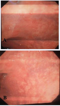
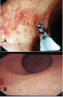
Subsequently we noticed that patients with colonic and rectal schistosomaisis especially in early presentation when doing endoscopy, they show elevated white dots with redness rim (figure 1(A, B)) or without redness rim (figure 2(A, B)) around usually in the rectum. When taking biopsy from such lesions we usually found schistosoma mansoni eggs.
Figure 1: Shows elevated white dots with redness rim around it.
Figure 2: Shows elevated white dots without red rim around it.
DISCUSSION
There is a consensus of pathological data that strongly support the association between S. japonicum and the colorectal cancer [7, 8]. In a review of the literature between 1898 and 1974, 276 cases of chistosomiasis japonicum associated with cancer of the large intestine were analysed by Chen et al. that support their previous results and giving better insight into the pathogenesis of schistosomal colorectal carcinoma. But, fewer of them revolve that S.mansoni has the same correlation with colonic cancer a study by Omer Salim, and in this review of the three cases could be an edition for this correlation.
The authors described presence of pseudopolyps, multiple ulcers, and hyperplastic ectopic submucosal glands, with evidence of oviposition and precancerous and cancerous transformation in these lesions with colonoscopic view of mucosal changes that appear as white dots surrounded by red rim (The Omer sign) this could has a great impact in early detection and moreover, early diagnosis and treatment.
In addition, it was demonstrated that the closer the area to the tumour the more ova tend to be detected Chen et al. A similar conclusion was drawn by Yu et al. from their studies on different types of schistosomal egg polyps [9,10].
While several case reports and descriptive studies have raised the possibilities of an association between S. mansoni infestation and colorectal malignancies, the pathological evidence supporting this claim is rather weak. But eliciting this newly invented sing could be a supportive evidence for diagnosis and further association.
The increase of the colorectal cancer among young population is found to be due to high rate of the chronic colitis which could be due to schistosomiasis in the areas of the endemicity [11].
CONCLUSION
Schistosomal colorectal carcinoma is an environmental disease that is mostly preventable by the early detection and implementation of screening strategies in the population at risk to establish early treatment that could prevent the occurrence of colorectal cancer. So, endoscopic detection of early schistosomiasis using The Omer sign could facilitate early detection of colorectal cancer.


