Novel Therapies for Neurodegenerative Diseases
- 1. Department of Medicine and Surgery, University of Benin, Nigeria
- 2. Department of Biochemistry, University of Port Harcourt, Nigeria
- 3. Department of Chemistry, University of Agriculture, Nigeria
- 4. Department of Medicine, University of Benin, Nigeria
Abstract
Background: Neurodegenerative diseases are becoming more common, which encourages biomedical scientists to research these illnesses. To tackle neurodegenerative disorders, new therapeutic techniques must be created to overcome the shortcomings of current therapeutic alternatives.
Methodology: This narrative review delves into the new approach to treating neurodegenerative diseases. A search of the English literature was conducted using the terms “gene therapy” together with “stem cell therapy,” “optogenetics,” “neurodegenerative diseases,” “novel approach,” and “treatment,” either independently or in combination, using PubMed, Scopus, and Research Gate.
Result: Conventional treatments for neurodegenerative disorders include pharmacotherapy, physical and occupational therapy, speech and swallowing therapy, and supportive care, but have limitations in cure, adverse effects, and long-term benefits. Advances in biomedical research are anticipated to positively impact neurological research, particularly in developing precise and novel therapy techniques, Contemporary approaches to neurodegenerative disease treatment have become more and more common in the domains of medicine and pharmacology in recent times. They comprise a number of state-of- the-art techniques, including RNA based therapy, optogenetics, gene therapy, immunotherapy, stem cell therapy, and non-invasive brain stimulation
Conclusion: The aging population is increasing neurodegenerative illnesses, necessitating novel therapeutic strategies like RNA based therapy, optogenetics, gene therapy, immunotherapy, stem cell therapy, and non-invasive brain stimulation. These personalized medicine approaches can improve understanding and identify affordable disease-modifying therapies for the healthcare system.
Keywords
• Novel Treatment; Neurodegenerative Disease; Gene Therapy; Optogenetics.
Citation
James E, Abigail O, Enenche NN, Ehizeme EN (2025) Novel Therapies for Neurodegenerative Diseases. J Neurol Transl Neurosci 10(1): 1101.
INTRODUCTION
As the population ages, neurodegenerative diseases like Alzheimer’s disease (AD), Parkinson’s disease (PD), multiple sclerosis (MS), amyotrophic lateral sclerosis (ALS), and Huntington’s disease (HD) are becoming more common, which encourages biomedical scientists to research these illnesses. Acute traumas can potentially worsen chronic neurodegenerative processes and raise systemic neuroinflammatory responses by impacting the central nervous system (CNS) [1]. Within the next 20 years, neurological illnesses are predicted by the World Health Organization (WHO) to overtake all other causes of death in humans. To tackle neurodegenerative disorders, new therapeutic techniques must be created to overcome the shortcomings of current therapeutic alternatives. The inability to find appropriate carriers for targeted medications keeps many sophisticated molecules from reaching their full potential [2]. Delays in brain and behavior are caused by a neurodegenerative disease, which causes neurons to gradually disappear and lose some of their functionality [3]. The non-neuronal cells of the central nervous system are called neuroglia or glial cells. The four forms of neuroglia that are involved in homeostasis, neuron protection, and brain plasticity regulation are oligodendrocytes, microglia, astrocytes, and NG2a-glia. Further harm to the central nervous system is caused by interactions between neurons, microglia, astrocytes, and oligodendrocytes, which leads to neuron and myelin loss [4]. The connection between neurodegenerative disorders and inflammation-related activated astrocytes has drawn more attention in recent years [5]. Numerous investigations have demonstrated a correlation between neurodegenerative illnesses and the prevalence of type 1 astrocytes, a neurotoxic phenotype that secretes proinflammatory cytokines. Moreover, in AD mice models, a greater quantity of activated microglia collaborated with abnormal astrocytes to reduce the survival of neurons [6 8]. It is generally recognized that dysfunctional microglia are crucial to neurodegeneration. Single-cell sequencing research in AD and ALS showed that as the disease advanced, the expression levels of numerous genes important for microglia function increased [9]. A variety of disorders are referred to as neurodegenerative illnesses, in which the brain or peripheral nervous system’s neurons gradually deteriorate. There aren’t many therapy choices for the majority of neurodegenerative disorders, such as Parkinson’s, Alzheimer’s, and Huntington’s, but there are some [10]. Currently, available techniques that can help control symptoms and reduce the progression of the disease include medications, physical and occupational therapy, speech therapy, assistive devices, and lifestyle modifications. It is important to realize that although these treatments alleviate symptoms and improve quality of life, the disease does not stop progressing or go back. Research into creating treatments that can alter the underlying degenerative processes and slow the advancement of these illnesses is ongoing [11]. This paper examines several cutting-edge methods, such as gene therapy, immunotherapy, stem cell therapy, optogenetics, RNA treatment, and non-invasive brain stimulation. Conventional Treatment for Neurodegenerative Disorders The main goals of conventional treatments for neurodegenerative illnesses are to control symptoms and reduce the rate at which the illness advances. The following are a few standard traditional treatments for neurodegenerative illnesses.
Pharmacotherapy: It is common practice to administer medication to treat the symptoms of neurodegenerative illnesses. To restore dopamine levels and reduce motor symptoms, for instance, levodopa is frequently prescribed for Parkinson’s disease. To improve cognitive performance in Alzheimer’s patients, cholinesterase inhibitors are utilized, such as donepezil. Medication is another option for treating symptoms like tremors, muscle stiffness, and sleep difficulties [12].
Physical and Occupational Therapy: Enhancing mobility, strength, balance, and coordination in individuals with neurodegenerative disorders can be facilitated by physical and occupational therapy. To support the maintenance of functional independence and quality of life, these therapies may incorporate stretches, exercises, gait training, and assistive technology [13].
Lifestyle Modification: Overall well-being in neurodegenerative illnesses can be enhanced by leading a healthy lifestyle. A healthy diet, stress reduction methods,regular exercise, and enough sleep are a few examples of this. It might also be advantageous to keep up social contacts and partake in mentally challenging pursuits [14].
Speech and Swallowing Therapy: Speech and swallowing can be impacted by neurodegenerative disorders. Through the instruction of methods to strengthen vocal muscles and enhance articulation, speech therapy can assist those who struggle with speaking. Conversely, the goal of swallowing therapy is to preserve safe and effective swallowing by teaching ways to avoid aspiration [15].
Supportive Care: Providing non-medical care to people with neurodegenerative disorders is known as supportive care. It involves emotional support, counseling, education for patients and their caretakers, and help with everyday tasks. A network of information and assistance can also be obtained through community services and support organizations [16].
LIMITATIONS WITH THE CONVENTIONAL METHODS
Conventional treatments for neurodegenerative illnesses have shortcomings. These include the lack of curative potential, the substantial side effects linked with pharmaceutical treatments, the inability to alter the course of the disease, the non-individualized approach that ignores individual variations, the incomplete management of symptoms, and the limited efficacy in slowing the progression of the disease and offering long-term benefits. Ongoing research focuses on the creation of innovative therapeutics, including gene therapies, immunotherapies, targeted therapies, and stem cell-based treatments, in order to get around these restrictions. The goal of these developments is to treat neurodegenerative disorders with more potent and disease-modifying therapies [17,18].
Novel Therapies: Advances in biomedical research are anticipated to have a positive impact on neurological research, particularly in the area of developing more precise and novel therapy techniques. In the fields of medicine and therapy, contemporary methods of treating illnesses have gained popularity recently. Concerns about advanced technologies’ creation, use, and regulation are growing as they are developed, used in therapy, reviewed by regulators, and subject to post-approval monitoring [2] Figure 1.
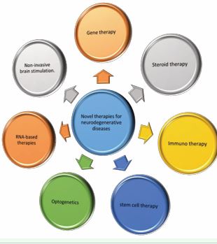
Figure 1 A schematic diagram showing the different novel therapeutic approaches to neurodegenerative diseases.
Gene Therapy: Neurodegenerative illnesses can be effectively treated by gene therapy, among other approaches. Developing successful therapeutic approaches against neurodegenerative illnesses requires an understanding of the fundamental mechanisms governing the control of gene expression at the spatial and temporal levels [19]. Nonetheless, one of the difficult parts of gene therapy by transduction is limiting leakage into adjacent tissues or perivascular spaces. To treat genetic abnormalities, produce therapeutic benefits, or alter biological functioning, therapeutic genes are delivered to target cells or tissues using gene therapy. To get therapeutic genes into target cells, a process known as transduction must occur. With MRI-guided convection enhanced delivery (iMRI-CED), real-time vector delivery monitoring has emerged as the gold standard for gene therapy [20]. If this cutting-edge neurosurgical approach is implemented successfully, promising preclinical therapeutics for neurodegenerative illnesses could be brought into clinical trials [21]. In gene therapy, a stable and inducible transgene is introduced to replace the defective gene with a controlled expression of the disease environment, so correcting the problem [22]. Because of the encouraging preliminary results, other investigators submitted protocols for phase I and II trials addressing a variety of neurological illnesses. Current advancements in clinical evaluation provide accurate knowledge of the anatomical-functional relationship, such as neuroimaging [23,24]. A plethora of preclinical and clinical research is required to demonstrate the efficacy of gene therapy in treating CNS illnesses [25,26]. It’s also important to remember that optimizing several aspects, including transgene selection, vector selection, and delivery mode selection, is necessary for the successful practice of gene therapy. The practice of gene therapy in neurodegenerative disease is difficult due to the intricacy of neural tissue and the interaction of the host immune system with vector and transgene. Furthermore, the application of gene therapy as a therapeutic approach presents a number of obstacles, including achieving the best possible therapeutic agent administration through intracerebral delivery, targeting growth factors, or directing therapeutic agents that stimulate the manufacture of growth factors into the brain. Parenchyma. Growth factors show promise in preclinical investigations, but they should be utilized cautiously. They also need to pass all stages of clinical trials. Growth factors, as opposed to other neuroprotective medicines, perform by restoring, protecting, and generating neurons along with their functionality through appropriate molecular pathways. Growth factors, although having a shorter half life, can still activate their corresponding receptors, which sets off a series of events that direct second messengers to activate transcription factors. The effects of this cascade can persist for days or months after growth factor inactivation [27,28].
Steroid Therapy: One potential preventive measure against neurodegenerative illnesses is improving neuronal function. Applying neuroactive and sex steroids can help improve neuronal function because they prevent neuronal death, oxidative stress, mitochondrial malfunction, and microglial activation, all of which are known to enhance survival, neurogenesis, and memory performance [29]. Based on the patient’s sex and neuropathology stage, these steroids may have favorable benefits. Furthermore, there is still a dearth of information regarding the use of steroids in the treatment of neurodegenerative illnesses. Indeed, there are several things to take into account before using steroids to treat neurodegenerative illnesses, such as formulation, dosage, administration route, bioavailability etc [30, 31].
Immunotherapy: The TNFα Targeting the synthesis of inflammatory mediators on the cell surface and in secretions that cause neuronal malfunction and death is a key piece of therapeutic strategy. In multiple animal models of neurodegenerative illnesses, such as AD and PD, inhibition of soluble TNFα using small-molecule inhibitors or viral over-expression has demonstrated efficacy [32,33]. Furthermore, weekly subcutaneous injections of the TNF inhibitor Etanercept did not enhance behavior, global function, or cognition in a small sample of people with mild-to-moderate AD dementia in a randomized double blind phase II clinical trial [34]. Given the availability of safe and efficacious TNFα inhibitors, which are presently employed to treat systemic inflammatory illnesses, they could be investigated at an earlier stage of the disease or in conjunction with additional anti-inflammatory remedies [32]. New studies investigating the potential anti inflammatory and immunomodulatory functions of molecules like glucagon-like peptide 1 (GLP-1), peroxisome proliferator-activated receptor gamma (PPAR-γ), and granulocyte-macrophage colony-stimulating factor (GM CSF) are being conducted in an effort to lessen the nature of neurodegenerative diseases [35].
A GM-CSF Deregulation of the immunomodulatory growth factor and cytokine GM-CSF is observed in neurodegenerative disorders. Actually, GM-CSF stimulates dendritic cells to regulate inflammation and microglial homeostasis by increasing microglial proliferation and inducing regulatory T (Treg) cell induction [36-38]. In preclinical and clinical investigations on AD, GM-CSF has demonstrated a broad range of neuroprotective potential. By saving hippocampal neuronal connections and improving Aβ clearance by attracting microglia to amyloid plaques, GM-CSF treatment effectively reduces neuroinflammation and cognitive deterioration in AD mice models [39, 40]. The 127-amino-acid synthetic recombinant form of GM-CSF, Sargamostim (GM-CSF Leukine), significantly improves cognitive and memory in groups treated with it when compared to controls, according to data from clinical trials. Furthermore, all AD patients have been found to be able to tolerate and safely use Sargamostim, according to phase 2 trials (NCT01409915) [41]. Furthermore, being studied in PD are sargamostim’s immunomodulatory and neuroprotective functions. According to preclinical and phase 1 trials (NCT01882010), PD-associated motor symptoms have significantly decreased, and GM-CSF has been demonstrated to rescue and protect nigrostriatal dopaminergic neurons through Treg induction [42,43]. These encouraging preliminary results support the notion that PD may not cause motor symptoms. On top of that, aside from modest injection site responses, sargamostim is safe and generally well-tolerated by patients [35].GLP-1 agonists lessen the amount of proinflammatory cytokines and the severity of the disease in mice models of AD and PD. There has been a thorough study of the mechanisms describing these medications’ modes of action and their routes [44]. However, recent data from GLP-1 agonist medication clinical trials for AD and PD has indicated minimal, if any, changes in disease pathogenesis [45,46]. Since these data are still preliminary, more research is necessary before appropriate inferences can be made [35]. According to preclinical research, PPAR-γ agonists improve motor performance and spatial memory in AD mice models by lowering neuroinflammation, Aβ 42 load, phosphorylated tau, and synaptophysin [47]. Moreover, T3D-959 (NCT04251182) and pioglitazone (NCT00982202), two highly promising PPAR-γ agonists, have demonstrated their safety and tolerability in patients during phase II clinical trials [48]. T3D-959 also markedly improved insulin metabolism and cognitive function in AD patients [48]. Clinical trials are still needed to confirm the preclinical effects of pioglitazone in Parkinson’s disease (PD), where it has significantly decreased neuroinflammation and microglial proliferation in several PD models [49]. Given the theory that misfolded proteins and Damage associated molecular patterns (DAMPs) regulate neuroinflammation by interacting with various Pattern recognition receptors (PRRs), PRRs like RAGE, Mac1, and Toll-like receptors (TLRs) are viable targets for treatment. Neuronal DAMP stimulation of PRRs has also been linked to the activation of downstream pro-inflammatory pathways, including NOX2, iNOS, and TNFα [35]. Therefore, one potential tactic to reduce reactive gliosis, persistent self perpetuating neuroinflammation, and neurotoxicity could be to effectively target PRRs implicated in each neurodegenerative illness. RAGE inhibitor PF-04494700 demonstrated encouraging outcomes in pre-clinical models; nevertheless, the outcomes of clinical trials yielded unclear results [50]. Using transgenic AD models, NLRP3 inhibitors—like MCC950—showed encouraging outcomes, demonstrating the significance of NLRP3 as an intracellular PRR that recognizes aggregates of misfolded proteins [35].
Stem Cell Therapy
Despite being relatively new, stem cell therapy has shown promise as a safe, interesting, and helpful treatment option for neurodegenerative illnesses [51]. For neurodegenerative diseases, the primary goal of stem cell therapy is to recreate a neural network that is comparable to the one that is destroyed due to the disease by obtaining particular neuronal subtypes [52]. Developing supplementary neuronal networks around afflicted areas or generating neurotrophic and scavenging toxic substances as a means of enhancing the environment to support host neurons is another strategy for treating neurodegenerative diseases [53]. Numerous clinical trials are looking into various facets of stem cell treatments for neurological conditions [54]. Thus far, the data appear to partially corroborate the findings from preclinical trials. Data, for instance, generally agree that the secretion of growth factors, such as nerve growth factor, glial cell line-derived neurotrophic factor, and brain-derived neurotrophic factor, achieves neuroprotection and is the basic mechanism underlying the observed improvements in neurodegenerative disorders [55]. Furthermore, a substantial body of research indicates that stem cell treatments can improve neurogenesis in individuals with neurodegenerative diseases [53]. the role of stem cell therapy in the treatment of some neurological diseases are explained below;
Stem Cell Therapy and Parkinson Disease: The available treatments for Parkinson’s disease (PD) include deep brain stimulation or medicines that raise dopamine (DA) levels by giving DA precursors such levodopa to make up for the DA shortfall brought on by the death of dopaminergic neurons [56]. While some existing drugs have shown promise in mitigating symptoms, they are unable to stop the disease’s progressive loss of dopaminergic neurons over time. On the other hand, a number of variables have been found to influence the development and course of the illness, such as oxidative stress, mitochondrial dysfunction, and abnormalities related to protein folding and the ubiquitin-proteasome system [57]. PD’s treatment is therefore complicated by the disease’s variety in pathology and underlying causes.Researchers have spent the last 20 years searching for different ways to supplement dopamine (DA) by growing stem-cell-derived dopaminergic neurons to replace those lost to the disease. In order to drive their development into the adult dopaminergic cell, scientists are utilizing Embryonic stem cells (ESCs), Neural stem cells (NSCs), and Induced Pluripotent Stem Cells (iPSCs) as stem cell typologies [58]. Animal models have shown some promise in the treatment of Parkinson’s disease (PD) using stem cell therapy. Brain cells extracted from human fetuses are being transplanted into patients with Parkinson’s disease (PD) as part of clinical trials to evaluate the procedure’s effectiveness while reducing potential negative effects. In the case of Parkinson’s disease patients, Schwarz et al. implanted dopaminergic neurons generated from human fetuses into their deficient striata [59]. Furthermore, ESCs have produced outstanding results in animal models because they can create dopaminergic neurons, something that has not been achieved with adult neural stem cells when treating Parkinson’s disease (PD). Furthermore, ESC migration into the parenchyma and spinal cord was demonstrated in rat models of spinal cord injury. Reversing motor deterioration in Parkinson’s disease (PD) is the ultimate goal, and they led to partial motor recovery. Although these experiments showed encouraging results at first, they were prohibited because of the possibility of cancer as well as ethical and religious issues [60]. NSCs have demonstrated encouraging outcomes in addition to ESCs. NSCs can release dopamine (DA) and hence reduce Parkinson’s disease symptoms since they exhibit a dopaminergic interneuron phenotype. In order to alleviate motor impairments in a PD rat model, Deleidi et al., reported that adult NSCs in the subventricular zone (SVZ) were successfully differentiated into functional midbrain dopaminergic neurons [61]. The symptoms of Parkinson’s disease were lessened in a rat model of the condition by Yasuhara et al.’s transplantation of human NSCs [62]. In a separate scenario, behavioral abnormalities in rats with Parkinson’s disease were corrected when nuclear-1-receptor-engineered NSCs from the SVZ developed into dopaminergic neurons [53].
Stem Cell Therapy and Alzheimer’s disease: Research suggests that by promoting neurogenesis, replacing lost neurons, and improving the environment, stem cell treatments may help to delay the progression of AD. It has been demonstrated that human NSCs are a promising source, as they have been shown to increase the expression of several cognition-related proteins in vivo in AD animal models, improve synaptic plasticity, lessen the burden of the pathology, and ameliorate spatial learning and memory impairment [63,64]. Notably, no decrease in tau or Aβ pathology was found, suggesting that while NSCs might regenerate, they cannot cure the underlying disease. Instead, they may assist balance the degenerative processes that occur in the AD brain [65]. By producing BDNF and enhancing cognitive performance, NSCs implanted in the hippocampus helped memory impairments in an AD animal model [66]. In addition, transplanted NSCs stimulate endogenous neural precursors, promote structural neuroplasticity, inhibit pro inflammatory cytokines, suppress neuronal apoptosis, and release growth factors. They also migrate and differentiate into different brain cells, including oligodendrocytes, astrocytes, and cholinergic neurons. Additionally, it was discovered by Kern et al., that NSCs transplanted into the hippocampal regions of elderly Down syndrome mice significantly reduce the number of tau/reelin-positive granules [67]. All things considered, it is yet unknown how NSCs encourage neurogenesis and cognitive function. Moreover, a barrier to treating AD with stem cell therapies is the creation of non-neuronal glial cell types through NSC transplantation [53]. It was recently discovered that using stem cells in combination with NGF could be an effective way to treat AD by inhibiting cell death, promoting the development of cholinergic neurons, and aiding in the creation of particular neuronal populations. NGF gene therapy has been researched in numerous animal models and has produced fruitful AD patient clinical studies [53].
Stem Cell Therapy and Huntington’s Disease: Therapeutic strategies based on stem cells have drawn a lot of interest as possible HD therapies. The replacement of lost or injured neurons and the alteration of mutant genes carrying enlarged CAG repeats are the two main goals of stem cell treatment for Huntington’s disease. Recent research indicates that NSCs have been the most often utilized stem cell type for treating HD. NSCs have been generated and extracted from a variety of sources, including HD patients’ somatic cells and the brain itself. There is strong evidence supporting the transplantation of stem cells or their derivatives in HD animal models, even if stem cell-based preclinical and clinical trials are still in their early stages. Early stem cell treatments using NSCs produced from ESCs and transplanted into HD animals have shown how motor neurons and circuit development are integrated in the host. Still, important questions remain about the moral and theological implications of fetal tissues [68]. The risks associated with stem cell treatments, such as non-neuronal cells inside grafts and graft overgrowth, should be taken into account. Mouse-derived NSCs function as GDNF delivery vehicles, acting favorably in lowering neuronal death and the ensuing motor dysfunction, according to a study by Ebert et al., on the brains of murine models of HD [69]. Engineered NPCs that overexpress GDNF were injected into HD rats in order to investigate the function of environmental enrichment in stem cell treatment for HD. While NSCs that had not been altered did not exhibit any neuroprotective properties, NPCs that expressed GDNF provided neuronal protection and functional recovery. Currently, Mesenchymal stem cells (MSCs) constitute a viable cell source for HD treatment due to their capacity to lower immune cell malfunction, increase compensatory neurogenesis, lower apoptosis, activate mitochondrial function, and encourage cell survival [53]. According to research published in 2010, Dey et al., found that in the YAC 128 mouse model of HD, MSCs that were genetically modified to overexpress BDNF or NGF reduced behavioral abnormalities and neuronal loss in the striatum [70]. That being said, the striatum may be able to slow down neurodegenerative processes by means of transplanting MSCs that overexpress BDNF [53]. The proliferation and neuronal differentiation of endogenous NSCs were found to be enhanced by Snyder et al., when they implanted human-derived MSCs into the dentate gyrus of the hippocampus in mouse models of HD [71]. Furthermore, Lin and colleagues demonstrated that human-derived MSCs provided neuroprotection and neurorestoration by means of neuronal differentiation, the capacity to provide neurotrophic support, and antiapoptotic functions. According to the findings, a mouse model of HD showed a marked decrease in motor impairment [72]. Since nearly all of the research has been done on animal models, stem cell therapy is still a long way from being used in a clinical setting to treat HD. More thorough, in-depth preclinical research will be required to verify its therapeutic potential.
Optogenetics
Combining optics and genetics to control individual neurons’ activity is known as optogenetics. Because of its precision in both space and time in regulating neural activity, it is a unique tool for neuroscience research. In optogenetics, light-sensitive opsins that either stimulate or inhibit neurons are the main component [73].
Optogenetics and Parkinson’s Disease: Optogenetics was applied in a recent study by Steinbeck and colleagues to explore graft function and graft to host connectivity [74]. Human embryonic stem cells (hESCs) were used to generate mesencephalic dopaminergic (mesDA) neurons, and the approach was used to modify the neurochemical and electrophysiological characteristics of these neurons [74]. Undifferentiated hESCs were transduced to produce either EYFP alone or halorhodopsin eNpHR3.0-EYFP (named HALO) under the direction of the human synapsin promoter in order to investigate the functionality of mesDA neurons implanted in lesioned striatum [75,76]. It was shown that motor impairments were created when HALO-expressing grafts were deactivated by light. Animals given apomorphine, an agonist for dopamine receptors D1 and D2, prior to deactivation of HALO-expressing grafts did not have a recurrence of motor impairments. Acute brain slice electrophysiological measurements showed that striatal GABA neurons’ excitatory postsynaptic potentials (EPSPs) and dopamine release from the graft were both triggered by stimulation of the corpus callosum [74]. On the other hand, elicited EPSP was significantly reduced by optogenetic silencing. This finding implies that the activation of D1 receptors by transplanted neurons enhances the EPSP response of host striatal GABA neurons. The results also point to the significance of graft neuronal activity and connection for Parkinson’s disease behavioral recovery [73].
Optogenetics and Hungtington’s Disease: A subset of medium-sized spiny neurons (MSNs) in mice models of HD has been found to have heightened spontaneous inhibitory synaptic activity, which attenuates striatal output. But it wasn’t apparent what was causing the higher inhibition. Utilizing optogenetic and electrophysiological techniques to evaluate feedforward and feedback inhibition in two transgenic mice models of HD, Cepeda and colleagues conducted a recent study to investigate the possible source(s) of enhanced inhibition [77]. GABAergic interneurons were specifically activated and the impact on GABA synaptic activity in MSNs was assessed by inserting channelrhodopsin-2 and EYFP into a double-floxed inverted open reading frame viral vector (AAV2-DIO-ChR2-EYFP) [77]. Two types of GABAergic interneurons, namely the fast-spiking (FS) and the persistent low-threshold spiking (PLTS) interneurons, were examined by single and dual patch-clamp recordings in MSNs of striatal slices. Under the direction of D1 (direct pathway) or D2 (indirect pathway) promoters, they saw specific changes in GABA synaptic activity in MSNs [78,79]. These results indicated that increased GABA activity on MSNs of the indirect route in HD is caused by several causes [77]. Feedforward inhibition from FS and PLTS interneurons provides the majority of the contribution. The bigger GABA-mediated synaptic responses originate from activated FS interneurons, whereas PLTS interneurons are in charge of the enhanced GABAergic spontaneous synaptic events. Nitric oxide (NO), neuropeptide Y (NPY), and somatostatin (SOM) are released by PLTS interneurons and may have neuroprotective properties [80,81]. Selective inhibition of FS and PLTS interneurons in the striatum may enhance the behavioral characteristics of HD. These results imply that altering the behavior of HD patients may benefit from selectively inhibiting the PLTS and FS interneurons.
Optogenetics and Alzheimer’s Disease: Alzheimer dementia (AD) patients’ brains have a characteristic pathological alteration known as amyloid beta peptide aggregation [82]. Nevertheless, the processes behind the release and accumulation of amyloid β peptides continued to be mysterious. Previous research utilizing pharmacological or electrical stimulations has demonstrated that neuronal Aβ production is activity dependent [83,84]. The precise paths implicated remained unclear, though. Optogenetics was used in a recent study by Yamamoto and colleagues to investigate the selective activation of a particular neural circuit in APP transgenic mice in order to observe the causal relationship between Aβ pathology and synaptic activation [73]. In the hippocampal perforant pathway of APP transgenic mice, stabilized step-function opsin (SSFO), a channel rhodopsin intended to induce a persistent neuronal hyperexcitability, was expressed. The CAMKIIα promoter-driven SSFO-EYFP adeno-associated virus vector was transduced unilaterally into the lateral entorhinal cortex with the specific aim of selectively stimulating the cortical projection neurons via the perforant pathway [73]. Following acute light stimulation, there was an approximately 24% rise in Aβ42 levels in the hippocampus interstitial fluid, as demonstrated by in vivo microdialysis. After five months of continuous optogenetic stimulation, Aβ deposits dramatically increased by a factor of 2.5 in mice. These results imply that increased Aβ deposition is linked to hyperactivity of a particular projection route. The groundwork for future studies employing optogenetics for long-term stimulation in animal models of neurodegenerative diseases was laid by this investigation [73].
RNA-based Therapies
For many of these neurodegenerative diseases, RNA based therapies offer an appealing therapeutic option because they allow for extremely precise regulation of gene expression, which can be used to either increase or decrease target protein levels depending on whether the mutation is causing a gain-of-toxic function or a loss-of function. RNA molecules are engineered to attach precisely to their target RNAs using Watson-Crick base pairing in order to facilitate RNA treatments. RNA treatments can thus be employed to specifically target important pathogenic pathways and/or biological origins of disease. One of the main benefits of RNA-based therapy is its ability to directly target any desired RNA, which significantly broadens the pool of potential targets beyond those that are typically druggable and available for small molecule- and antibody-based treatments. Antisense oligonucleotides (ASOs) and RNA interference (RNAi) are the two main kinds of RNA treatments. Single-stranded RNAs known as ASOs selectively attach to and modify target RNAs to affect translation or turnover, whereas double-stranded RNA molecules are used by RNAi to silence genes [85].
The potential of RNA-based therapies as a personalized medicine modality is enormous. Targeting particular genes and possibly even patient mutations is possible with RNA therapies due to their versatility. Designing ASOs to downregulate protein levels (e.g., SOD1, huntingtin, and tau) is the most widely used strategy when it comes to ASOs. When it comes to focusing on essential cellular pathways and proteins involved in the pathophysiology of disease, as well as mutant proteins with gain-of-toxic function, this kind of mechanism is very helpful. ASOs can also be developed to modify splicing of the target RNA (such as for the SMN2 RNA by nusinersen) to successfully enhance the quantities of functional SMN protein [85]. Furthermore, by blocking the nonsense-mediated mRNA decay process,ASOs can be engineered to prevent the degradation of mutant mRNAs and block microRNAs. We go into greater depth about each of these various ASO-based strategies in relation to particular neurodegenerative illnesses below [85].Reducing protein expression to minimize the quantities of hazardous aggregates has been one of the most popular applications for ASOs. To do this, specific mRNA transcript degradation is induced, as seen in the cases of tau, huntingtin, and SOD1. The oligonucleotides are made to bind to the coding mRNA transcripts and cause RNA: DNA hybrids to be specifically broken down by the enzyme ribonuclease (RNase) H1. Five to seven core DNA nucleotides are used in the creation of ASOs in this instance. The first gene linked to hereditary ALS, superoxide dismutase 1 (SOD1), has been effectively targeted with this strategy. 20% of cases of familial ALS are caused by mutations in SOD1. In 20% of cases of familial ALS, the mutation is in SOD1 [86]. The majority of the evidence points to a gain-of-toxic function rather than a loss of function as the mechanism by which SOD1 mutations cause disease, albeit this is still uncertain. Once successful in preclinical studies, ASOs intended to lower SOD1 levels proceeded to clinical trials for testing. For neurodegenerative illness, this was the first application of ASOs. Clinical trials for the medication Tofersen are currently in phase III [87,88]. All of these studies demonstrated the effectiveness of targeted therapy in distributing ASOs throughout the central nervous system. This treatment was safe, corresponded with a meaningful drop in SOD1 levels, and reduced patient illness. Research on RNA-based therapeutics in the setting of several neurodegenerative illnesses is now being conducted by a number of laboratories within the Henry and Amelia Nasrallah Center for Neuroscience. Targeting important proteins implicated in the pathophysiology of AD with ASOs has been the focus of Susan Farr’s group. Research conducted on mice models of AD has shown that ASOs that reduce levels of presenilin-1, GSK-3β, and amyloid precursor protein (APP) enhance memory and learning [89,90]. Her lab has been researching traumatic brain injury (TBI) more recently, and they have shown that post-injury injection of ASOs targeting GSK-3β protects cognitive deficits [91]. Based on these data, it is highly likely that ASOs that target these proteins can benefit cognitively in individuals with TBI and AD in a similar way.
Non-invasive brain stimulation
For years, it has been suggested that non-invasive brain stimulation (NIBS) techniques, such as transcranial direct current stimulation (tDCS), transcranial alternating current stimulation (tACS), and repetitive transcranial magnetic stimulation (rTMS), can alleviate both motor and non-motor symptoms in a variety of neurological conditions, including neurodegenerative disorders like Alzheimer’s disease (AD) and Parkinson’s disease (PD) [92,93]. For the purpose of modifying cortical and, most likely, subcortical activity, they are secure and effective tools [94].
Non-invasive brain stimulation and Parkinson’s Disease: Numerous neuromodulation methods have been proposed, including complementary therapy approaches that are non-invasive [95], and invasive [96]. Transcranial direct current stimulation (tDCS) has emerged as a promising therapeutic option that has been shown to improve motor and cognitive functions [97]. Nevertheless, there is still much to learn about the processes by which tDCS affects PD patients, especially at the cellular and molecular levels [98]. According to available data, tDCS causes an increase in dopamine release [99,100], modifies alpha-synuclein aggregation and autophagic degradation [98], modifies the concentration of neurotransmitters (such as glutamate, serotonin, and GABA) [101], and has anti-inflammatory and anti-apoptotic effects. These cellular effects, however, have only been observed in animal models or in vitro; human studies have not yet verified them. Under these circumstances, it is anticipated that tDCS will upregulate the expression of brain-derived neurotrophic factor (BDNF) [102], a neurotrophic factor and neurotransmitter modulator that promotes neurogenesis [103] and neuronal survival. Based on animal studies [104], these results imply a potential neuroprotective impact of tDCS, which seems to be partially efficient in repairing some of the metabolic abnormalities associated with neurodegenerative disorders. In fact, what is known now about neurotoxin-treated animal models reveals encouraging initial findings about the antioxidant function caused by tDCS and the ability of dopaminergic cells to withstand neurotoxin-induced cell death [105].
Non-invasive brain stimulation and Alzheimer’s Disease: For the treatment of cognitive symptoms linked to AD, tDCS or rTMS have been used in a number of randomized clinical trials [106]. Neurotrophic factors (NTFs) have been thoroughly investigated in relation to AD. NTFs control the growth, survival, proliferation, migration, and differentiation of neurons [107]. The temporal cortex and hippocampus are among the damaged brain regions in AD where decreased expression of NTFs, such as nerve growth factor (NGF), BDNF, glial cell line-derived neurotrophic factor (GDNF), and ciliary neurotrophic factor (CNTF), has been noted. The beneficial effect of neuromodulation techniques on brain-derived neurotrophic factor (BDNF) has recently attracted special attention. BDNF is one of the most significant cellular mechanisms underlying learning and memory and is required in the hippocampus for late-phase long-term potentiation. Furthermore, by promoting the development and survival of cholinergic neurons in the basal forebrain, BDNF stimulates the release of acetylcholine [108]. Notably, compared to controls, several investigations have recently revealed elevated BDNF levels following rTMS in the basal forebrain and hippocampus in AD animal models, as well as in the serum of AD patients [109]. On NGF brain levels, rTMS was also found to be beneficial. Additionally, the BDNF-TrkB signaling pathway [110], which influences cell survival, migration, axon and dendritic outgrowth, synaptogenesis, synaptic transmission, and synapse remodelling [111], was also positively affected by rTMS and tDCS.
FUTURE PERSPECTIVES
In this discussion, we covered a few cutting-edge molecular targets that are being investigated and may prove to be useful for upcoming therapeutic approaches treating Neurodegenerative diseases. Due to differential expressions in microglial-specific receptors (CD33, TREM2, and CR3), genetic alterations in pathways linked to immune inflammatory mediated responses have been shown to be significant risk factors for late-onset AD (LOAD) [112]. It has been recently determined that tyrosine kinase binding protein (TYROBP), a key microglial transmembrane signaling adaptor polypeptide that is a direct adaptor for TREM2, CD33, and CR3 activity and may possess an AD related function. The dysregulation of both variables and TYROBP function may be genetically associated hazards to AD. TREM2 activity is influenced by both factors. In AD transgenic animals, however, a reduction in microglial activation surrounding the plaques was linked to TYROBP deficiency. An interesting new treatment strategy for AD may be revealed by the observed change in microglial autophagy flux [113]. Additionally, in Parkinson’s disease (PD), focusing on chaperone-mediated autophagy constituents such lysosome-associated membrane protein 2a (LAMP2a) and Hsc70 may be an alternate strategy to promote autophagic flux. Increasing LAMP2a in vitro in the SH-SY5Y DA cell line, rat primary cortical neurons, and nigral DA neurons in vivo has been shown to reduce α-Syn aggregation and toxicity, protecting these neurons [113].Extracellular vesicles (EVs) produced from mesenchymal stem cells (MSCs) have been used in recent advances in the treatment of neurological illnesses. The MSCs secrete these EVs, which are membrane-bound, tiny structures that contain a variety of bioactive substances, including lipids, proteins, and nucleic acids. It has been found by researchers that these EVs have therapeutic qualities and can have positive impacts on the nervous system. EVs’ capacity to penetrate the blood-brain barrier and reach the brain’s damaged areas is a major benefit of employing them to treat neurodegenerative diseases [2]. Neuronal survival, neurogenesis (the creation of new neurons), and neuroinflammation are all improved by EVs produced from MSCs, which slows the progression of neurodegenerative disorders. Furthermore, certain molecules, such microRNAs, which are essential for controlling gene expression, can be transferred by these EVs. EVs have the ability to modify gene expression patterns and enhance neuroprotection by transferring these microRNAs to specific brain cells. Additionally, therapeutic medications or compounds can be delivered to specific brain regions with the help of EVs produced from MSCs [2].
In the field of regenerative medicine, there is a growing interest in the utilization of extracellular vesicles (EVs) produced from mesenchymal stem cells (MSCs). Proteins, lipids, and nucleic acids are only a few of the bioactive substances found in MSC-EVs, which are tiny membrane bound vesicles secreted by MSCs. The following are some potential directions for MSC-EV therapy in the future:
Targeted Delivery: By modifying their genetic makeup, MSC-EVs can be made to specifically transport therapeutic materials like microRNA, growth hormones, or small interfering RNA (siRNA). Targeting certain cells or tissues impacted by neurodegenerative illnesses is possible with these cargo-loaded MSC-EVs. It may be possible to improve the stability, prevent degradation, and improve the distribution of therapeutic compounds to the intended site of action by encasing them in EVs [2].
Immunomodulation: MSC-EVs are identical to their parent cells in that they have immunomodulatory capabilities. They have the ability to control inflammation, alter immunological responses, and encourage tissue healing. With immunomodulatory effects comparable to those of cell-based therapies, MSC-EVs hold promise as a cell-free substitute for MSC transplantation [2].
Combination Therapy: To increase the efficacy of other therapy, MSC-EVs can be administered in conjunction with them. MSC-EVs, for instance, can be utilized in conjunction with gene therapy techniques or given in addition to existing neuroprotective medications. The results of treatment may be enhanced by this synergistic combination’s additive or even synergistic effects [2].
CONCLUSION
Today’s aging population is contributing to the rising incidence of neurodegenerative illnesses, which impact a vast number of people. Treatments available at this time are only symptomatic. In order to stop or reduce the development of these ailments, it is imperative to discover novel therapeutic strategies. In this instance, we examined innovative methods including RNA based therapy, optogenetics, gene therapy, immunotherapy, stem cell therapy, and non-invasive brain stimulation. Depending on the patient’s disease progression and the type of disease (idiopathic or familial), personalized medicine based on single- or multi-target drug approaches including gene therapy and RNA based therapy may be helpful in improving understanding and identifying disease-modifying therapies that are affordable for the healthcare system.
STATEMENTS AND DECLARATIONS
Author Contributions
Emmanuel James came up with the research topic and wrote the introduction, conventional therapies for neurodegenerative diseases and its shortcomings, novel therapies including gene therapy, stem cell therapy, optogenetics and immunotherapy for management of neurodegenerative diseases.
Onoja Abigail contributed to RNA based therapy for neurodegenerative diseases and also contributed to the compilation of the manuscript.
Ngbede Nelson Enenche contributed to conventional therapy and steroid therapy for treatment of neurodegenerative diseases and the restructuring of the manuscript.
All authors contributed equally to the research
All authors read and approved the final manuscript.
REFERENCES
- Holbrook JA, Jarosz-Griffiths HH, Caseley E, Lara-Reyna S, Poulter JA, Williams-Gray CH, et al. Neurodegenerative disease and the NLRP3 inflammasome. Front Pharmacol. 2021; 12: 643254.
- Palanisamy CP, Pei J, Alugoju P, Anthikapalli NVA, Jayaraman S, Veeraraghavan VP, et al. New strategies of neurodegenerative disease treatment with extracellular vesicles (EVs) derived from mesenchymal stem cells (MSCs). Theranostics. 2023; 13: 4138-4165.
- Ullah MF, Ahmad A, Bhat SH, Abu-Duhier FM, Barreto GE, Ashraf GM. Impact of sex differences and gender specificity on behavioral characteristics and pathophysiology of neurodegenerative disorders. Neurosci Biobehav Rev. 2019; 102: 95-105.
- Weigel M, Wang L, Fu Mm. Microtubule organization and dynamicsin oligodendrocytes, astrocytes, and microglia. Dev Neurobiol. 2021; 81: 310-320.
- Scarfò G, Piccarducci R, Daniele S, Franzoni F, Martini C. Exploring the Role of Lipid-Binding Proteins and Oxidative Stress in Neurodegenerative Disorders: A Focus on the Neuroprotective Effects of Nutraceutical Supplementation and Physical Exercise. Antioxidants. 2022; 11: 2116.
- Taylor X, Cisternas P, Jury N, Martinez P, Huang X, You Y, et al. Activated endothelial cells induce a distinct type of astrocytic reactivity. Commun Biol. 2022; 5: 282.
- Jiang X, Li S, Feng X, Li L, Hao J, Wang D, et al. Mushroom polysaccharides as potential candidates for alleviating neurodegenerative diseases. Nutrients. 2022; 14: 4833.
- Radenovic L, Nenadic M, U?amek-Kozio? M, Januszewski S, Czuczwar SJ, Andjus PR, et al. Heterogeneity in brain distribution of activated microglia and astrocytes in a rat ischemic model of Alzheimer’s disease after 2 years of survival. Aging. 2020; 12: 12251.
- Olah M, Menon V, Habib N, Taga MF, Ma Y, Yung CJ, et al. Single cell RNA sequencing of human microglia uncovers a subset associated with Alzheimer’s disease. Nat Commun. 2020; 11: 6129.
- Baloni P, Funk CC, Readhead B, Price ND. Systems modeling of metabolic dysregulation in neurodegenerative diseases. Curr Opin Pharmacol. 2021; 60: 59-65.
- Armstrong MJ, Okun MS. Diagnosis and treatment of Parkinson’s disease: a review. JAMA. 2020; 323: 548-560.
- Drijgers RL, Aalten P, Winogrodzka A, Verhey FR, Leentjens AF. Pharmacological treatment of apathy in neurodegenerative diseases: a systematic review. Dement Geriatr Cogn Disord. 2009; 28: 13-22.
- Arbesman M, Lieberman D, Berlanstein DR. Method for the systematic reviews on occupational therapy and neurodegenerative diseases. Am J Occup Ther. 2014; 68: 15-19.
- Santiago JA, Potashkin JA. Physical activity and lifestyle modifications in the treatment of neurodegenerative diseases. Front Aging Neurosci. 2023; 15: 1185671.
- Tye CB, Gardner PA, Dion GR, Simpson CB, Dominguez LM. Impact of fiberoptic endoscopic evaluation of swallowing outcomes and dysphagia management in neurodegenerative diseases. Laryngoscope. 2021; 131: 726-730.
- Armitage A, Fonkem E. Supportive care of neurodegenerative patients. Front Oncol. 2023; 13: 1029938.
- Ramanathan S, Archunan G, Sivakumar M, Selvan ST, Fred AL, Kumar S, et al. Theranostic applications of nanoparticles in neurodegenerative disorders. Int J Nanomed. 2018; 13: 5561-5576.
- Vissers MF, Heuberger JA, Groeneveld GJ. Targeting for success: demonstrating proof-of-concept with mechanistic early phase clinical pharmacology studies for disease-modification in neurodegenerative disorders. Int J Mol Sci. 2021; 22: 1615.
- D’Souza GX, Rose SE, Knupp A, Nicholson DA, Keene CD, Young JE. The application of in vitro-derived human neurons in neurodegenerative disease modeling. J Neurosci Res. 2021; 99: 124-140.
- Richardson RM, Larson P. Direct Convective Nervous System Drug Delivery for Patients with Neurodegenerative Disorders. Nervous System Drug Delivery: Elsevier. 2019: 463-472.
- Rosser AE, Busse ME, Gray WP, Badin RA, Perrier AL, Wheelock V, et al. Translating cell therapies for neurodegenerative diseases: Huntington’s disease as a model disorder. Brain. 2022; 145: 1584- 1597.
- Richardson RM, Varenika V, Forsayeth JR, Bankiewicz KS. Future applications: gene therapy. Neurosurg Clin N Am. 2009; 20: 205-210.
- Van Horn JD, Pelphrey KA. Neuroimaging of the developing brain. Brain Imaging Behav. 2015; 9: 1–4.
- Deverman BE, Ravina BM, Bankiewicz KS, Paul SM, Sah DW. Gene therapy for neurological disorders: progress and prospects. Nat Rev Drug Discov. 2018; 17: 641-659.
- Simonato M, Bennett J, Boulis NM, Castro MG, Fink DJ, Goins WF, et al. Progress in gene therapy for neurological disorders. Nat Rev Neurol. 2013; 9: 277-291.
- Ginn SL, Amaya AK, Alexander IE, Edelstein M, Abedi MR. Gene Therapy clinical trials worldwide to 2017: an update. J Gene Med. 2018; 20: e3015.
- Sudhakar V, Richardson RM. Gene therapy for neurodegenerative diseases. Neurother J Am Soc Exp Neurother. 2019; 16: 166-175.
- Sidorova YA, Saarma M. Can growth factors cure Parkinson’s disease? Trends Pharmacol Sci. 2020; 41: 909-922.
- Akwa Y. Steroids and Alzheimer’s disease: changes associated with pathology and therapeutic potential. Int J Mol Sci. 2020; 21: 4812.
- Deeb O, Nabulsi M. Exploring Multiple Sclerosis (MS) and Amyotrophic Lateral Scler osis (ALS) as neurodegenerative diseases and their treatments: a review study. Curr Top Med Chem. 2020; 20: 2391-2403.
- Renaud J, Martinoli MG. Considerations for the use of polyphenols as therapies in neurodegenerative diseases. Int J Mol Sci. 2019; 20: 1883.
- Barnum CJ, Chen X, Chung J, Chang J, Williams M, Grigoryan N, et al. Peripheral administration of the selective inhibitor of soluble tumor necrosis factor (TNF) XPro® 1595 attenuates nigral cell loss and glial activation in 6-OHDA hemiparkinsonian rats. J Parkinson Dis. 2014; 4: 349-360.
- Detrait ER, Danis B, Lamberty Y, Foerch P. Peripheral administration of an anti-TNF-alpha receptor fusion protein counteracts the amyloid induced elevation of hippocampal TNF-alpha levels and memory deficits in mice. Neurochem Int. 2014; 72: 10-13.
- Butchart J, Brook L, Hopkins V, Teeling J, Puntener U, Culliford D, et al. Etanercept in Alzheimer disease: a randomized, placebo-controlled, double-blind, phase 2 trial. Neurology. 2015; 84: 2161-2168.
- Mortada I, Farah R, Nabha S, Ojcius DM, Fares Y, Almawi WY, et al. Immunotherapies for Neurodegenerative Diseases. Front Neurol. 2021; 12: 654739.
- Bhattacharya P, Budnick I, Singh M, Thiruppathi M, Alharshawi K, Elshabrawy H, et al. Dual role of GM-CSF as a pro-inflammatory and a regulatory cytokine: implications for immune therapy. J Interfer Cytok Res. 2015; 35: 585-599.
- Dikmen HO, Hemmerich M, Lewen A, Hollnagel JO, Chausse B, KannO. GM-CSF induces noninflammatory proliferation of microglia and disturbs electrical neuronal network rhythms in situ. J Neuroinflammation. 2020; 17: 235.
- Esen N, Kielian T. Effects of low dose GM-CSF on microglial inflammatory profiles to diverse pathogen-associated molecular patterns (PAMPs). J Neuroinflammation. 2007; 4: 10.
- Kiyota T, Machhi J, Lu Y, Dyavarshetty B, Nemati M, Yokoyama I, et al. Granulocyte-macrophage colony-stimulating factor neuroprotective activities in Alzheimer’s disease mice. J Neuroimmunol. 2018; 319: 80-92.
- Sanchez-Ramos J, Song S, Sava V, Catlow B, Lin X, Mori T, et al. Granulocyte colony stimulating factor decreases brain amyloidburden and reverses cognitive impairment in Alzheimer’s mice. Neuroscience. 2009; 163: 55-72.
- Potter H, Woodcock JH, Boyd TD, Coughlan CM, O’Shaughnessy JR, Borges MT, et al. Safety and efficacy of sargramostim (GM-CSF) in the treatment of Alzheimer’s disease. Alzheimers Dement (N Y). 2021; 7: e12158.
- Gendelman HE, Zhang Y, Santamaria P, Olson KE, Schutt CR, Bhatti D, et al. Evaluation of the safety and immunomodulatory effects of sargramostim in a randomized, double-blind phase 1 clinical Parkinson’s disease trial. NPJ Parkinsons Dis. 2017; 310.
- Kosloski LM, Kosmacek EA, Olson KE, Mosley RL, Gendelman HE. GM-CSF induces neuroprotective and anti-inflammatory responses in 1-methyl-4-phenyl-1,2,3,6-tetrahydropyridine intoxicated mice. J Neuroimmunol. 2013; 265: 1-10.
- Glotfelty EJ, Olson L, Karlsson TE, Li Y, Greig NH. Glucagon-like peptide-1 (GLP-1)-based receptor agonists as a treatment for Parkinson’s disease. Expert Opin Investig Drugs. 2020; 29: 595-602.
- Gejl M, Gjedde A, Egefjord L, Møller A, Hansen SB, Vang K, et al. In Alzheimer’s Disease, 6-Month Treatment with GLP-1 Analog Prevents Decline of Brain Glucose Metabolism: Randomized, Placebo- Controlled, Double-Blind Clinical Trial. Front Aging Neurosci. 2016; 8: 108.
- Watson KT, Wroolie TE, Tong G, Foland-Ross LC, Frangou S, Singh M, et al. Neural correlates of liraglutide effects in persons at risk for Alzheimer’s disease. Behav Brain Res. 2019; 356: 271-278.
- Tong M, Deochand C, Didsbury J, de la Monte SM. T3D-959: A Multi- Faceted Disease Remedial Drug Candidate for the Treatment of Alzheimer’s Disease. J Alzheimers Dis. 2016; 51: 123-138.
- Chamberlain S, Gabriel H, Strittmatter W, Didsbury J. An Exploratory Phase IIa Study of the PPAR delta/gamma Agonist T3D-959 Assessing Metabolic and Cognitive Function in Subjects with Mild to Moderate Alzheimer’s Disease. J Alzheimers Dis. 2020; 73: 1085-1103.
- Machado MMF, Bassani TB, Cóppola-Segovia V, Moura ELR, Zanata SM, Andreatini R, et al. PPAR-γ agonist pioglitazone reduces microglial proliferation and NF-κB activation in the substantia nigra in the 6-hydroxydopamine model of Parkinson’s disease. Pharmacol Rep. 2019; 71: 556-564.
- Sabbagh MN, Agro A, Bell J, Aisen PS, Schweizer E, Galasko D. PF- 04494700, an oral inhibitor of receptor for advanced glycation end products (RAGE), in Alzheimer disease. Alzheimer Dis Assoc Disord. 2011; 25: 206-212.
- Karussis D, Petrou P, Kassis I. Clinical experience with stem cells and other cell therapies in neurological diseases. J Neurol Sci. 2013; 324: 1-9.
- Lunn JS, Sakowski SA, Hur J, Feldman EL. Stem cell technology for neurodegenerative diseases. Ann Neurol. 2011; 70: 353-361.
- Sivandzade F, Cucullo L. Regenerative Stem Cell Therapy for Neurodegenerative Diseases: An Overview. Int J Mol Sci. 2021; 22: 2153.
- Nicaise C, Mitrecic D, Falnikar A, Lepore AC. Transplantation of stem cell-derived astrocytes for the treatment of amyotrophic lateral sclerosis and spinal cord injury. World J Stem Cells. 2015; 7: 380-398.
- Fu MH, Li CL, Lin HL, Chen PC, Calkins MJ, Chang YF, et al. Stem cell transplantation therapy in Parkinson’s disease. Springerplus. 2015; 4: 597.
- Raza C, Anjum R, Shakeel NUA. Parkinson’s disease: Mechanisms, translational models and management strategies. Life Sci. 2019; 226: 77-90.
- Trombetta-Lima M, Sabogal-Guáqueta AM, Dolga AM. Mitochondrial dysfunction in neurodegenerative diseases: A focus on iPSC-derived neuronal models. Cell Calcium. 2021; 94: 102362.
- Kim HJ. Stem cell potential in Parkinson’s disease and molecular factors for the generation of dopamine neurons. Biochim Biophys Acta. 2011; 1812: 1-11.
- Schwarz J, Storch A. Transplantation in Parkinson’s disease: will mesenchymal stem cells help to reenter the clinical arena? Transl Res. 2010; 155: 55-56.
- Tom CM, Younesi S, Meer E, Bresee C, Godoy M, Mattis VB. Survival of iPSC-derived grafts within the striatum of immunodeficient mice: Importance of developmental stage of both transplant and host recipient. Exp Neurol. 2017; 297: 118-128.
- Deleidi M, Cooper O, Hargus G, Levy A, Isacson O. Oct4-induced reprogramming is required for adult brain neural stem cell differentiation into midbrain dopaminergic neurons. PLoS One. 2011; 6: e19926.
- Yasuhara T, Matsukawa N, Hara K, Yu G, Xu L, Maki M, et al. Transplantation of human neural stem cells exerts neuroprotection in a rat model of Parkinson’s disease. J Neurosci. 2006; 26: 12497- 12511.
- Banigan MG, Kao PF, Kozubek JA, Winslow AR, Medina J, Costa J, et al. Differential expression of exosomal microRNAs in prefrontal cortices of schizophrenia and bipolar disorder patients. PLoS One. 2013; 8: e48814.
- Yeo RW, Lai RC, Zhang B, Tan SS, Yin Y, Teh BJ, et al. Mesenchymal stem cell: an efficient mass producer of exosomes for drug delivery. Adv Drug Deliv Rev. 2013; 65: 336-341.
- Ager RR, Davis JL, Agazaryan A, Benavente F, Poon WW, LaFerla FM, et al. Human neural stem cells improve cognition and promote synaptic growth in two complementary transgenic models of Alzheimer’s disease and neuronal loss. Hippocampus. 2015; 25: 813-826.
- Sakthiswary R, Raymond AA. Stem cell therapy in neurodegenerative diseases: From principles to practice. Neural Regen Res. 2012; 7: 1822-1831.
- Kern DS, Maclean KN, Jiang H, Synder EY, Sladek JR Jr, Bjugstad KB. Neural stem cells reduce hippocampal tau and reelin accumulation in aged Ts65Dn Down syndrome mice. Cell Transplant. 2011; 20: 371- 379.
- Connor B. Concise Review: The Use of Stem Cells for Understanding and Treating Huntington’s Disease. Stem Cells. 2018; 36: 146-160.
- Ebert AD, Barber AE, Heins BM, Svendsen CN. Ex vivo delivery of GDNF maintains motor function and prevents neuronal loss in a transgenic mouse model of Huntington’s disease. Exp Neurol. 2010; 224: 155-162.
- Dey ND, Bombard MC, Roland BP, Davidson S, Lu M, Rossignol J, et al. Genetically engineered mesenchymal stem cells reduce behavioral deficits in the YAC 128 mouse model of Huntington’s disease. Behav Brain Res. 2010; 214: 193-200.
- Snyder BR, Chiu AM, Prockop DJ, Chan AW. Human multipotent stromal cells (MSCs) increase neurogenesis and decrease atrophy of the striatum in a transgenic mouse model for Huntington’s disease. PLoS One. 2010; 5: e9347.
- Lin YT, Chern Y, Shen CK, Wen HL, Chang YC, Li H, et al. Human mesenchymal stem cells prolong survival and ameliorate motor deficit through trophic support in Huntington’s disease mouse models. PLoS One. 2011; 6: e22924.
- Vann KT, Xiong ZG. Optogenetics for neurodegenerative diseases. Int J Physiol Pathophysiol Pharmacol. 2016; 8: 1-8.
- Steinbeck JA, Choi SJ, Mrejeru A, Ganat Y, Deisseroth K, Sulzer D, et al. Optogenetics enables functional analysis of human embryonic stem cell-derived grafts in a Parkinson’s disease model. Nat Biotechnol. 2015; 33: 204-209.
- Kriks S, Shim JW, Piao J, Ganat YM, Wakeman DR, Xie Z, et al. Dopamine neurons derived from human ES cells efficiently engraft in animal models of Parkinson’s disease. Nature. 2011; 480: 547-551.
- Kirkeby A, Grealish S, Wolf DA, Nelander J, Wood J, Lundblad M, et al. Generation of regionally specified neural progenitors and functional neurons from human embryonic stem cells under defined conditions. Cell Rep. 2012; 1: 703-714.
- Cepeda C, Galvan L, Holley SM, Rao SP, André VM, Botelho EP, et al. Multiple sources of striatal inhibition are differentially affected in Huntington’s disease mouse models. J Neurosci. 2013; 33: 7393-7406.
- Cepeda C, André VM, Yamazaki I, Wu N, Kleiman-Weiner M, Levine MS. Differential electrophysiological properties of dopamine D1 and D2 receptor-containing striatal medium-sized spiny neurons. Eur J Neurosci. 2008; 27: 671-682.
- André VM, Cepeda C, Fisher YE, Huynh M, Bardakjian N, Singh S, et al. Differential electrophysiological changes in striatal output neurons in Huntington’s disease. J Neurosci. 2011; 31: 1170-1182.
- Kawaguchi Y, Wilson CJ, Augood SJ, Emson PC. Striatal interneurones: chemical, physiological and morphological characterization. Trends Neurosci. 1995; 18: 527-535.
- Deckel AW. Nitric oxide and nitric oxide synthase in Huntington’s disease. J Neurosci Res. 2001; 64: 99-107.
- Selkoe D, Mandelkow E, Holtzman D. Deciphering Alzheimer disease. Cold Spring Harb Perspect Med. 2012; 2: a011460.
- Kamenetz F, Tomita T, Hsieh H, Seabrook G, Borchelt D, Iwatsubo T, et al. APP processing and synaptic function. Neuron. 2003; 37: 925- 937.
- Cirrito JR, Yamada KA, Finn MB, Sloviter RS, Bales KR, May PC, et al. Synaptic activity regulates interstitial fluid amyloid-beta levels in vivo. Neuron. 2005; 48: 913-922.
- Ayala YM, Nguyen AD. RNA-Based Therapies for Neurodegenerative Diseases. Mo Med. 2021; 118: 340-345.
- Rosen DR, Siddique T, Patterson D, Figlewicz DA, Sapp P, Hentati A, et al. Mutations in Cu/Zn superoxide dismutase gene are associated with familial amyotrophic lateral sclerosis. Nature. 1993; 362: 59-62.
- McCampbell A, Cole T, Wegener AJ, Tomassy GS, Setnicka A, Farley BJ, et al. Antisense oligonucleotides extend survival and reverse decrement in muscle response in ALS models. J Clin Invest. 2018; 128: 3558-3567.
- Miller T, Cudkowicz M, Shaw PJ, Andersen PM, Atassi N, Bucelli RC, et al. Phase 1-2 Trial of Antisense Oligonucleotide Tofersen for SOD1 ALS. N Engl J Med. 2020; 383: 109-119.
- Armbrecht HJ, Siddiqui AM, Green M, Farr SA, Kumar VB, Banks WA, et al. Antisense against Amyloid-β Protein Precursor Reverses Memory Deficits and Alters Gene Expression in Neurotropic and Insulin-Signaling Pathways in SAMP8 Mice. J Alzheimers Dis. 2015; 46: 535-548.
- Farr SA, Sandoval KE, Niehoff ML, Witt KA, Kumar VB, Morley JE. Peripheral Administration of GSK-3β Antisense Oligonucleotide Improves Learning and Memory in SAMP8 and Tg2576 Mouse Models of Alzheimer’s Disease. J Alzheimers Dis. 2016; 54: 1339-1348.
- Farr SA, Niehoff ML, Kumar VB, Roby DA, Morley JE. Inhibition ofGlycogen Synthase Kinase 3β as a Treatment for the Prevention ofCognitive Deficits after a Traumatic Brain Injury. J Neurotrauma.2019; 36: 1869-1875.
- Ferrucci R, Mameli F, Guidi I, Mrakic-Sposta S, Vergari M, Marceglia S, et al. Transcranial direct current stimulation improves recognition memory in Alzheimer disease. Neurology. 2008; 71: 493-498.
- Lefaucheur JP, Antal A, Ayache SS, Benninger DH, Brunelin J, Cogiamanian F, et al. Evidence-based guidelines on the therapeutic use of transcranial direct current stimulation (tDCS). Clin Neurophysiol. 2017; 128: 56-92.
- Guidetti M, Arlotti M, Bocci T, Bianchi AM, Parazzini M, Ferrucci R, et al. Electric Fields Induced in the Brain by Transcranial Electric Stimulation: A Review of In Vivo Recordings. Biomed. 2022; 10: 2333.
- Guidetti M, Marceglia S, Loh A, Harmsen IE, Meoni S, Foffani G, et al. Clinical perspectives of adaptive deep brain stimulation. Brain Stimul. 2021; 14: 1238-1247.
- Madrid J, Benninger DH. Non-invasive brain stimulation for Parkinson’s disease: Clinical evidence, latest concepts and future goals: A systematic review. J Neurosci Methods. 2021; 347: 108957.
- Simpson MW, Mak M. The effect of transcranial direct current stimulation on upper limb motor performance in Parkinson’s disease: a systematic review. J Neurol. 2020; 267: 3479-3488.
- Sala G, Bocci T, Borzì V, Parazzini M, Priori A, Ferrarese C. Direct current stimulation enhances neuronal alpha-synuclein degradation in vitro. Sci Rep. 2021; 11: 2197.
- Fonteneau C, Redoute J, Haesebaert F, Le Bars D, Costes N, Suaud- Chagny MF, et al. Frontal Transcranial Direct Current Stimulation Induces Dopamine Release in the Ventral Striatum in Human. Cereb Cortex. 2018; 28: 2636-2646.
- Tanaka T, Takano Y, Tanaka S, Hironaka N, Kobayashi K, Hanakawa T, et al. Transcranial direct-current stimulation increases extracellular dopamine levels in the rat striatum. Front Syst Neurosci. 2013; 7: 6.
- Fritsch B, Reis J, Martinowich K, Schambra HM, Ji Y, Cohen LG, et al. Direct current stimulation promotes BDNF-dependent synaptic plasticity: potential implications for motor learning. Neuron. 2010; 66: 198-204.
- Ranieri F, Podda MV, Riccardi E, Frisullo G, Dileone M, Profice P, et al. Modulation of LTP at rat hippocampal CA3-CA1 synapses by direct current stimulation. J Neurophysiol. 2012; 107: 1868-1880.
- Benraiss A, Chmielnicki E, Lerner K, Roh D, Goldman SA. Adenoviral brain-derived neurotrophic factor induces both neostriatal andolfactory neuronal recruitment from endogenous progenitor cells in the adult forebrain. J Neurosci. 2001; 21: 6718-6731.
- Lee SB, Youn J, Jang W, Yang HO. Neuroprotective effect of anodal transcranial direct current stimulation on 1-methyl-4-phenyl- 1,2,3,6-tetrahydropyridine (MPTP)-induced neurotoxicity in mice through modulating mitochondrial dynamics. Neurochem Int. 2019; 129: 104491.
- Feng XJ, Huang YT, Huang YZ, Kuo CW, Peng CW, Rotenberg A, et al. Early transcranial direct current stimulation treatment exerts neuroprotective effects on 6-OHDA-induced Parkinsonism in rats. Brain Stimul. 2020; 13: 655-663.
- Rajji TK. Transcranial Magnetic and Electrical Stimulation in Alzheimer’s Disease and Mild Cognitive Impairment: A Review of Randomized Controlled Trials. Clin Pharmacol Ther. 2019; 106: 776-780.
- Xiao N, Le QT. Neurotrophic Factors and Their Potential Applications in Tissue Regeneration. Arch Immunol Ther Exp (Warsz). 2016; 64: 89-99.
- Knipper M, da Penha Berzaghi M, Blöchl A, Breer H, Thoenen H, Lindholm D. Positive feedback between acetylcholine and the neurotrophins nerve growth factor and brain-derived neurotrophic factor in the rat hippocampus. Eur J Neurosci. 1994; 6: 668-671.
- Velioglu HA, Hanoglu L, Bayraktaroglu Z, Toprak G, Guler EM, Bektay MY, et al. Left lateral parietal rTMS improves cognition and modulates resting brain connectivity in patients with Alzheimer’s disease: Possible role of BDNF and oxidative stress. Neurobiol Learn Mem. 2021; 180: 107410.
- Chen X, Dong GY, Wang LX. High-frequency transcranial magnetic stimulation protects APP/PS1 mice against Alzheimer’s disease progress by reducing APOE and enhancing autophagy. Brain Behav. 2020; 10: e01740.
- Ohira K, Hayashi M. A new aspect of the TrkB signaling pathway in neural plasticity. Curr Neuropharmacol. 2009; 7: 276-285.
- Haure-Mirande JV, Audrain M, Fanutza T, Kim SH, Klein WL, Glabe C, et al. Deficiency of TYROBP, an adapter protein for TREM2 and CR3 receptors, is neuroprotective in a mouse model of early Alzheimer’s pathology. Acta Neuropathol. 2017; 134: 769-788.
- Xilouri M, Brekk OR, Landeck N, Pitychoutis PM, Papasilekas T, Papadopoulou-Daifoti Z, et al. Boosting chaperone-mediated autophagy in vivo mitigates α-synuclein-induced neurodegeneration. Brain. 2013; 136: 2130-2146.


