Loading

Journal of Neurology and Translational Neuroscience

What Immunopathogenic Similarities Exist Between SARSCov-2 Infection And Multiple Sclerosis?: A Plea For Daily Vitamin D Supplementation To Improve Quality Of Life!

Research Article | Open Access | Volume 8 | Issue 1

  • 1. Independent researcher, Internal medicine, rehabilitation, social medicine Hochwaldstrasse 2 D-97769 Bad Brückenau, Germany
+ Show More - Show Less
Corresponding Authors
Hans-Klaus Goischke, Internal medicine, rehabilitation, social medicine Hochwaldstrasse 2 D-97769 Bad Brückenau, Germany, Bavaria
Abstract

For decades, arguments have been made for vitamin D supplementation as an add-on therapy “for” and “against” for people with multiple sclerosis. The COVID-19 pandemic has challenged therapists to protect high-risk groups, such as people with multiple sclerosis, from further health damage. In addition to the risk factors established in the general population, such as older age, male gender, hypertension, endocrinological and cardiovascular diseases, advanced disability, highly active disease with frequent drug therapies and doctor consultations as well as comorbidities accumulate in MS.

COVID-19 and MS have overlapping clinical and immunopathogenic properties. Due to the immunomodulatory properties of VitD, adjunctive therapy is likely to be consistent when COVID-19 occurs in PwMS.


Keywords

• Multiple sclerosis

• Immunopathogenesis

• SARS-CoV-2 infection

• Ebstein-Barr virus

• Vitamin D supplementation

• Ocrelizumab

CITATION

Goischke HK (2023) What Immunopathogenic Similarities Exist Between SARS-Cov-2 Infection And Multiple Sclerosis?: A Plea For Daily Vitamin D Supplementation To Improve Quality Of Life!. J Neurol Transl Neurosci 8(1): 1093.

INTRODUCTION

Multiple sclerosis (MS) is an inflammatory neurodegenerative disease with a suspected autoimmune origin. The disease begins earlier than current diagnostic criteria can detect. It affects the entire central nervous system and not just the white matter, as the original term – inflammatory demyelinating disorder suggests [1]. It is characterized by a very heterogeneous course of the disease, which is represented by relapse-associated neurological deterioration, but also by an increase in disability that is independent of relapse [2]. MS is associated with reduced vitamin D status [3].

Vitamin D (VitD) (1, 25(OH) 2D3) a neurosteroid, is known to influence brain development, neutransmission, and to exert neuroprotective and immunomodulatory functions [4,5].

The major form of vitamin D (Vit D), 25(OH) D and its active hormonal form, 1, 25-dihydroxy-vitamin D3, are present in the brain [6-9].

VitD signal transduction occurs via the vitamin D receptor (VDR) [5]. VDR is a part of the nuclear receptor superfamily of transcription factors [4]. Translocation of VDR to the nucleus is enhanced by calcitriol [10].

The VDR is expressed in almost all cell types of the body and various brain regions as well as in immune cells [11,12]. VDR is present in numerous brain cells such as oligodendrocytes, astrocytes, microglia and neurons [6]. The enzymes that convert 25(OH) D to 1, 25(OH) 2D3 (CYP27B1) and the enzymes responsible for its degradation (CYP24A1) have been detected by immunohistochemical methods [4]. In the human brain, VDR have been found in the hippocampus, hypothalamus, amygdala, cortex, cerebellum and substantia nigra, [6,12,13].

Although the cause of multiple sclerosis (MS) is not yet fully understood , interactions between B and T cells are discussed in the pathogenesis , such as peripheral escape of B cells from T cell-mediated control , interaction of pathogenic B and T cells in secondary lymph nodes and reactivation of B and T cells accumulating in the CNS (details in [14]. Auto reactive inflammatory cells , including effector T cells (Th1, Th17, CD8 + cytotoxic T cells), activated B cells and plasma cells producing autoantibodies infiltrates the central nervous system (CNS) [15].

This T cell migration into the CNS is the key to the pathogenesis of MS. The Treg cells (CD4+CD25+Foxp3 regulatory cells [Treg]) play an essential role in this. A dysfunction of both the Treg cells and the Tr1 (IL-10-secreting regulatory type 1 cells) has been confirmed [16,17]. Subsets of B cells function as antigen presenting cells and pro- inflammatory cytokine producing cells [18].

A supplementary supportive therapy to improve the course of the disease in people with multiple sclerosis (PwMS) is daily vitamin D supplementation with the aim of achieving s25(OH)D values of at least 60-80ng/ mL (30-130ng/ mL ) [19,20].

The effect of vitamin D (VitD) on the innate and acquired immune system is undisputed and 1, 25-dihydroxyvitamin D plays a major role in cell homeostasis in the CNS. 1, 25(OH) 2 D3 is autocrine and paracrine [21].

1, 25(OH) 2D3 also targets macrophages, monocytes, dendritic cells (DC), and T and B cells. VitD regulates the activation of microglia and astrocytes [22,23].

In DC, it decreases IL-12, TNF alpha and IL-6 production and promotes IL-10 production. The active metabolites of VitD also promotes the development of forkhead box protein 3 (FOXP3+ regulatory T (Treg) cells and IL-10-producing T regulatory type 1 (TR1) cells. 1, 25(OH) 2D3 blocks B- cell proliferation, plasma cell differentiation and immunoglobulin production [24-26].

Vitamin D exerts its complex immunomodulatory effects on CD4+ T lymphocytes (Th1, Th2, Th17, Treg cells), among others, and the secret of pro- inflammatory cytokines (IL-2, IL-6, IL-12, IL- 17, IFN-γ, TNF-α and TNF-β) is inhibited. It stimulates the production of anti- inflammatory regulatory Th2 cytokines (IL-4, IL-5 and IL-10) [26-29].

The cytokine profiles is shifted from a Th1 to a Th2 mediated profile (decrease in IFN-gamma and increase in IL-4 [28]. As a result, vitamin D insufficiency in PwMS must be avoided at all costs [28,30].

Not only is vit D deficiency associated with MS risk, but s25 (OH) D levels are inversely correlated with risk of relapse, CNS lesions, and disability progression. Vit D suppl. reduces the number of new Gd +- enhancing or new / enlarged T2 lesions on MRI [28-31].

COVID-19 infection

Severe acute respiratory syndrome coronavirus 2 (SARS CoV-2) is an exceptionally transmissible and pathogenic coronavirus that emerged in late 2019 and caused an acute respiratory disease pandemic known as coronavirus infection 2019 (COVID-19). New omicron variants are constantly being discovered [32].

COVID-19 can develop into a severe disease associated with immediate and delayed sequelae in various organs, including the central nervous system (CNS) [32].

Over the last 3 years, a complex connection between SARS CoV-2 infection and MS has emerged [33].

The renewed risk of a serious infection from COVID-19 should be an additional motivation for daily high-dose vitamin D administration [34,35]. In the context of prevention, it is worth mentioning that with circulating s25 (OH) D values ≥ 55ng/ mL, the SARS-CoV-2 positivity rate was significantly lower than with values below or with deficiency [36]. Current data suggest a protective role of VitD, particularly with a lower risk of intensive care unit admission and a reduced risk of death [37, 38]. In addition, the occurrence of long-COVID is an aspect of implementing this simple, effective, safe and cost-effective therapy with a broad therapeutic window for the prevention and treatment of COVID-19 disease [39-41].

Although there is still no indisputable evidence that Vit D supplementation (VitD suppl.) reduces the risk of SARS-Cov-2 infection in healthy people, but there is collective evidence that at-risk people have a benefit [42]. PwMS with comorbidities, psychiatric illnesses, hypertension, obesity (an increased BMI may correlate with a severe course of Covid-19), age > 50 years, severe disability and methylprednisolone boost therapy as well as some DMTs (disease- modifying therapies ) have a higher risk of infection and an increased risk of severe COVID-19 courses [43-45]. Infections (SARS-COV-2) can increase MS symptoms (pseudo-relapses) or cause real relapses [46]. In post-COVID syndrome (long COVID), one in eight patients presents with symptoms such as fatigue, shortness of breath, cough, joint pain, chest pain, muscle pain, headache and paresthesia in the limbs after at least 3 months. The latter can also occur in PwMS per se [47,48].

Mechanisms of action of Vit D on Covid-19

There have been hundreds of publications on this in the years since the outbreak of the COVID-19 infection. If people at risk have VitD supply. There is a lower risk of infection, the severity of the disease with admission to the intensive care unit or a reduced risk of death, Long Covid occurs less frequently [37-41,49-52]. It is not ethically justifiable to withhold a high dose of VitD from people at risk.

Barrea et al. list in detail 14 mechanisms as described by Vit D supply. The risk of COVID-19 infection can be reduced and sufficient Covid 19 vaccination is supported [48,53-58]. Vit D and its metabolites inactivate viruses (increase the antimicrobial peptide cathelicidin , lead to reduction of the risk of a cytokine storm , reduce matrix metalloproteinase-9 concentration and thereby increase the host’s metabolic tolerance to damage, reduce the risk of pneumonia and myocarditis, lead to the reduction of the concentration of pro-inflammatory cytokines, in particular interleukin 6 (IL-6), which promotes the permeability of the BBB, which leads to the potentiation of CNS damage in PwMS, is serious [48,60- 62].

Vit D enables neuroprotection by reducing inflammation and oxidative stress. Low 25(OH) D levels were inversely correlated with high IL-6 levels and were independent predictors of COVID-19 severity and mortality [63]. 1, 25(OH) 2D3 inhibits immunoglobulin synthesis, regulates B cell activity and reduces auto-Ab production. It converts B cells into plasma cells [37]. VitD reduces the risk of infection with EBV [48].

Long COVID- 19, Epstein-Barr virus (EBV), MS and inflammation

Current studies show evidence that the chronic inflammation in long Covid-19 infection with a reactivation of the latent Epstein-Barr virus (EBV) can lead to a worsening of the health status in PwMS [64-68]. In MS, there is a high affinity molecular mimicry between the EBV transcription factor EBNA-1 and the CNS protein GlialCAM (glial cell adhesion molecule of the central nervous system [69]. Bernal et al were able to detect EBV reactivation by detecting EBV DNA and antibodies against EBV lytic Genes provide [70].

In 66.7% of Long Covid patients, EBV reactivation could be demonstrated by a positive titer for EBV EA-D (early antigen- diffuse). IgG or EBV-VCA (viral capsid antigen)-IgM) can be provided [65]. Long COVID patients with fatigue and neurocognitive disorders were associated with serological evidence of recent EBV reactivation (early antigen D [EA-D] IgG positivity) or high nuclear antigen IgG levels [71].

The triad of inflammatory markers IL-1ß, IL-6 and TNF can be found in both Long Covid and MS [72-74]. Low sun exposure acts synergistically with high EBNA-1 Ab levels and was associated with an increased risk of MS [75].

There is a connection between high EBNA-1 antibody levels and low s25 (OH) D levels. On the other hand, a high dose of VitD lowers. The EBNA-1 antibody levels in PwMS [76-78]. Another parallel arises from the increase in GFAP (glial fibrillary acidic protein) as a functional disorder of the astrocytes around 4 months after the start of SARS-Cov2 infection [71]. The concentration of NfL (Neurofilament light chain), GFAP and total tau in CSF in patients with COVID-19 was often elevated with neurological symptoms [79]. Elevated sNfL has already been verified in mild to moderate COVID-19 disease [80]. On the other hand, the risk of mortality increased if sNfL and sGFAP levels were already elevated upon hospital admission [81].

Because there are no effective drugs that block EBV reactivation in Long Covid [82], there are multiple arguments for Vit D suppl. High-dose Vit D- Suppl (14,000IU/ day) for 48 weeks or 20,000 IU/week for 48 weeks selectively reduced anti-EBNA-1 antibody levels in PwMS (RRMS) [78,83].

Several mechanisms are under discussion: 1. VitD could induce better clearance of EBV-infected B cells, 2. Vit D could directly target and impair viral replication in EBV-infected cells, 3. Produce better control of inflammation in general, 4 In an EBV mediated inflammatory cascade, 1,25(OH)D3 could suppress the activation of reactive astrocytes 5. It is likely that at high s25 (OH) D levels, the VitD receptor EBNA 2 (Epstein -Barr virus nuclear antigen 2) is displaced upon DNA binding [78,83-88].

Early start of therapy is a crucial factor

The early start of therapy for VitD suppl. Is crucial in influencing influenza and COVID-19 infections [89].

The Corona-19 mortality risk therefore correlates inversely with the VitD status and a mortality rate close to zero could theoretically be achieved at over 50ng/ mL s25(OH)D [90]. The importance of Vit-D metabolism as a potential prophylactic, immunoregulatory and neuroprotective treatment of infections (COVID-19) is increasingly being considered in clinical practice as part of a multitherapeutic approach [91-93].

Dosage suggestions for vitamin D supplementation

Currently, there are no consensus guidelines suggesting an appropriate concentration of serum 25(OH) D to prevent COVID-19 or reduce its morbidity and mortality.

It is becoming increasingly clear to start with a “ loading dose ” with high doses of VitD over a few days and then continue with a “maintenance dose”, although various variants have been put up for discussion.

For example, one study used a weekly or fortnightly dose totaling 100,000–200,000 IU over 8 weeks (1800 or 3600 IU/ day) [94]. To obtain 75nmol/l s25 (OH) D values, the following equation was described:

Dose (IU) = 40 x (75 – serum 25(OH) D (3) [nmo /L] x body weight [94].

Over 30ng/ mL s25 (OH) D values were also achieved with a single oral dose of 200,000–600,000 IU [58, 95].

An s25 (OH) D level of 40-60ng/ml could be achieved by dosing up to 6000IU/day for several weeks [19,96]. A daily VitD intake of 10,000IU/day for 4 weeks would lead to a faster optimal s25 (OH) D level in the “status nascendi” of an infection [38].

Another dosage regimen was recommended: cholecalciferol 0.532 mg on day 1 and continued with 0.266 mg on days 3,7,14,21,28 (1 IU vitamin D3 ? 0.025 µ g vitamin D3 ? 65.0 mmol vitamin D3 [97]. The pharmacokinetic properties of calcifediol allow rapid absorption within hours, facilitating the immediate availability of 25OHD3 in the target tissue. This has drastically reduced the need for intensive care unit admission and the mortality rate [97]. Continued daily VitD dose will depend on the s25 (OH) values. The “maintenance dose” depends very individually on the achieved 25(OH) D serum levels and could be 4000-5000 IU/day (dose-response effect) [98].

Serum D level >50 ng / mL with a daily intake of 5000 IU (4000-7000 IU) and with obesity , gastrointestinal absorption disorders, antiepileptic or retroviral therapy, a two to four times higher dosage of VitD is required [99].

Daily doses of vitamin D have a therapeutic advantage over bolus doses

Reducing pro-inflammatory cytokines requires daily VitD administration because the cytokines have a short half-life of a few minutes to hours and are continuously produced by immune cells to remain elevated [100-102]. A daily VitD administration results in a better overall protective effect on general respiratory tract infections than a bolus administration [103].

Tores et al. was able to detect an increase in the anti inflammatory cytokine IL-10 in COVID-19 infection by administering 10,000IU/day for 14 days and achieve a shorter hospital stay compared to those patients who only received 2000IU/day [104]. Why very high doses of VitD supplementation may be necessary also arises from the knowledge that the SARS Cov-2 virus downregulates the VitD receptors, so that only high doses can still have an effect [48]. [Barrera]. Furthermore, VitD modulates ACE2 (Angiotensin Converting Enzyme 2) and can thereby cause ARDS (Acute Respiratory Distress syndrome) and alleviate acute lung damage caused by SARS-Cov-2 [105,106].

The dosage depends on the time and severity of the illness

However, for severe respiratory infections (COVID-19) or sepsis, a single oral bolus dose (100,000 to 500,000 IU) or in a divided dose of several times 50,000 IU is administered to ensure a rapid, adequate intracellular supply of calcitrol within 3 to 5 days and to make it available [107]. The COVIT-TRIAL study showed that in at-risk elderly patients, early administration of high-dose VitD in a single dose of 400,000IU within 72 hours of diagnosis improved all-cause mortality on day 14 compared to a standard dose of 50,000IU [49] . Short-term cholecalciferol administration with 60,000 IU over 7 days led to negative SARS-Cov-2 RNA in 41.7% of patients with mild symptoms or asymptomatic [108].

DISCUSSION

Both diseases, COVID-19 and MS, are associated with dysregulation of the immune system and mutually affect the course of the disease (pathophysiological/immunological mechanisms in detail in [32, 109]. The occurrence and severity of Covid-19 disease are associated with a worsening of clinical disability PwMS is linked, with several hypotheses currently being discussed. Details in [110]. In a prospective, ongoing MRI study (3 Tesla) (Post-Hospitalization COVID-19 Study) over 5-6 months, a reduction in the volume of the gray area was shown substance, white matter hyperintensities and vascular injury patterns. These qualitative MRI abnormalities were observed in 50% of post-COVID patients [111].

Elevated CRP (≥5mg/ mL) was associated with regional brain atrophy, requiring preventive measures through VitD suppl. challenges [112-114].

Low 25(OH)D levels are associated with a higher risk of infection, COVID-19 severity, respiratory distress severity, length of hospital stay, and mortality, and acute sequelae (long COVID) occur more frequently [92,115-119].

Before the previous consistent evidence of a connection between low s25(OH) values and negative SARS-Cov2 results in the general population is confirmed in high-quality, randomized studies [36], the administration of Vit D should be used for pathobiological reasons in PwMS .

The COVID-19 infection reinforces the need for a concept of a strategic MS treatment approach using all available therapies based on scientific knowledge and current experience [120].

Since the beginning of the coronavirus disease (COVID- 19) pandemic, different national recommendations for relapse therapy in RRMS with glucocorticoids as well as risk assessment statements have been issued for the management of PwMS [121,122]. And an international consensus Statement on care in the pandemic and post-pandemic period published in 2021 [123].

In the following years it became clear that patient-related data, such as increasing age, higher BMI, male gender, activity and type of MS subtypes, higher disability (higher EDSS), type of drug therapy, such as anti-CD -20 DMTs ( Ocrelizumab ) play a relevant role in optimizing and minimizing risk in the care of PwMS [124-126]. Janzuel et al reported that the use of anti CD-20 therapies in PwMS and RRMS was associated with severe COVID-19 courses [127]. On the other hand, it has been shown that for younger, more often female people with active disease and further therapies, the risk of infection increased through contact with doctors and medical facilities [128].

Vitamin D - a modifiable parameter in COVID-19 infection

It is undisputed in the literature that vitamin D exerts immunoregulatory properties on both the innate and the adaptive immune system and its deficiency is associated with increased autoimmunity and susceptibility to infections [28,32,96,129,130]. If in the general population a high s25(OH)D level (>30mg/ mL ) is associated with a lower risk of SARS-CoV-2 infection and severe COVID-19 progression as well as lower mortality compared to people with VitD insufficiency [129,131], daily VitD is recommended for PwMS suppl. On [48,96,118,131 136].

If the s25(OH)D value is recognized as a modifying factor for the positive influence of COVID-19 and LONG-COVID in PwMS , appropriate information must be given to this vulnerable group of people [99]. Those PwMS who have undergone anti-CD-20 therapy require special attention because severe COVID-19 courses are to be expected.

PwMS and ocrelizumab therapy showed breakthrough infection rates of 59% after the third SARS-CoV-2 mRNA vaccination [137]. Prophylactic VitD administration would therefore be particularly recommended to protect PwMS from infections. Although there are uncertainties regarding the optimal daily dosage of VitD (dose-response relationship taking body weight into account), the negative consequences could be reduced by pushing back the global “pandemic of VitD insufficiency/deficiency” [138-141]. In the era of COVID-19 infection and long-COVID [118,142-148].

COVID-19 and the endocrine system

SARS-CoV-2 penetrates the host cell primarily via ACE2 and TMPRSS2 (Tran’s membrane protease Serine2). These major receptors are also expressed in endocrine organs (thyroid, adrenal, hypothalamus, pituitary, gonads, and pancreas) [149,150]. During an infection of the endocrine system, the SARS-CoV-2 virus RNA has been detected in all endocrine organs, including the thyroid [149,151]. SARS-Cov-2 antigen was confirmed histopathologically in the thyroid gland postmortem [152].

Interactions between SARS-CoV-2 infection and the thyroid were confirmed by the description of “SARS-CoV-2-associated thyroiditis” in patients with COVID-19 [153]. An increase in thyroid autoantibodies TPOAb (thyroid peroxidase antibody) and TgAb (thyroglobulin-antibody) were observed during and after Covid-19 infection [154,155]. The prevalence of autoimmune thyroid diseases in the post-Covid phase was doubled compared to a control group [155]. People with a pre-existing autoimmune disease and Covid-19 were 23% more likely to be diagnosed with another autoimmune disease [156].

PwMS are affected by an increased manifestation of a coexisting autoimmune thyroid disease, especially Hashimoto thyroiditis [157]. This group of people with comorbidities such as Hashimoto’s thyroiditis or Graves’ disease should therefore be monitored endocrinologically both during the acute SARS CoV-2 infection and in the post-acute phase. Thyroid dysfunction is the most common comorbidity during therapy with DMT alemtuzumab [158].

In long-term follow-up, new symptoms that arise could easily be identified as being caused by endocrine disorders [159]. The exact mechanisms are not yet fully understood [45,150]. VitD deficiency could promote the autoimmune process [160,161]. A VitD supply. Lowers the serum TPO-Ab and TgAb titers and therefore this add-on therapy should also be used for prevention, especially when the endocrine system is involved [143,150,162 165].

Gut-microbiota-brain axis

Overlaps between Covid-19 and MS can be seen in the involvement of the gut-microbiota-brain axis. Dysfunctionality is characterized by changes in the bacterial composition and diversity of the microbiota [29,32,166-168]. A high-dose VitD supply. Could provide a benefit [41,168-170].

CONCLUSION

The clinical use of vitamin D supplementation will play an important role in the prevention and control of COVID-19 disease. Most research proved that vitamin D supplementation would bring clinical benefits to COVID-19 patients in terms of prevention and treatment (hospital length, mortality, improvement in blood oxygen saturation, better prognosis) [171].

There are no specific recommendations for everyday practice in connection with COVID-19 and vitamin D supplementation in patients with multiple sclerosis, but the scientific community also regrets that despite numerous “pro” and “against” voices, no consensus has been reached [172]. New corona variants will emerge, and, for ethical reasons, it is not justifiable to wait many years for the results of ongoing large-scale randomized studies [143] and their implementation in practice will certainly be delayed for years. Perhaps the idea is widely held that in a non existent ideal world all health decisions can only be made on the basis of overwhelming evidence, but a time of crisis may require slightly different rules [172].

The fight against SARS-CoV-2 is far from over because further virus variants will emerge. Eliminating vitamin D deficiency quickly is easy, inexpensive and quick to do. Depending on the course of the disease, comorbidities, treatment with disease modifying therapies and other diverse factors, multiple sclerosis is associated with an increased risk of a possible severe course of the COVID-19 infection. Vitamin D supplementation to prevent and influence the course of a COVID-19 infection in PwMS is presented and the supplementation protocols used to date are listed from the literature.
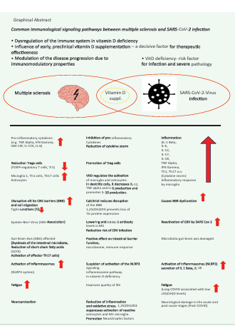
Graphical Abstract

Graphical Abstract

REFERENCES

1. Heming M, Wiendl H. Learning multiple sclerosis immunopathogenesis from anti-CD20 therapy. Proc Natl Acad Sci USA. 2023; 120: e2221544120

2. Lublin FD, Häring DA, Ganjgahi H, Ocampo A, Hatami F, ?uklina J, et al. How patients with multiple sclerosis acquire disability. Brain. 2022; 145: 3147-3161.

3. Rolf L, Damoiseaux J, Huitinga I, Kimenai D, van den Ouweland J, Hupperts R, et al. Stress-Axis Regulation by Vitamin D3 in Multiple Sclerosis. Front Neurol. 2018; 9: 263.

4. Cui X, Eyles DW. Vitamin D and the Central Nervous System: Causative and Preventative Mechanisms in Brain Disorders. Nutrients. 2022; 14: 4353.

5. Cui X, Gooch H, Petty A, McGrath JJ, Eyles D. Vitamin D and the brain: Genomic and non-genomic actions. Mol Cell Endocrinol. 2017; 453:131-143.

6. Eyles DW. Vitamin D: Brain and Behavior. JBMR Plus. 2020; 5: e10419.

7. Eyles DW, Smith S, Kinobe R, Hewison M, McGrath JJ. Distribution of the vitamin D receptor and 1 alpha-hydroxylase in human brain. J Chem Neuroanat. 2005; 29: 21-30.

8. Holmøy T, Moen SM. Assessing vitamin D in the central nervous system. Acta Neurol Scand Suppl. 2010; (190):88-92.

9. Fu X, Dolnikowski GG, Patterson WB, Dawson-Hughes B, Zheng T, Morris MC,et al. Determination of Vitamin D and Its Metabolites in Human Brain Using an Ultra-Pressure LC-Tandem Mass Spectra Method. Curr Dev Nutr. 2019; 3: nzz074.

10. Laso? W, Jantas D, Le?kiewicz M, Regulska M, Basta-Kaim A. The Vitamin D Receptor as a Potential Target for the Treatment of Age-Related Neurodegenerative Diseases Such as Alzheimer’s and Parkinson’s Diseases: A Narrative Review. Cells 2023, 12, 660.

11. Pistono C, Osera C, Monti MC, Boiocchi C, Mallucci G, Cuccia M, et al. Vitamin D Receptor and Its Influence on Multiple Sclerosis Risk and Severity: From Gene Polymorphisms to Protein Expression. Immuno 2022; 2: 469-481.

12. Gombash SE, Lee PW, Sawdai E, Lovett-Racke AE. Vitamin D as a Risk Factor for Multiple Sclerosis: Immunoregulatory or Neuroprotective? Front Neurol. 2022; 13: 796933.

13. Liu H, He Y, Beck J, da Silva Teixeira S, Harrison K, Xu Y, et al. Defining vitamin D receptor expression in the brain using a novel VDRCre mouse. J Comp Neurol. 2021; 529: 2362-2375.

14. van Langelaar J, Rijvers L, Smolders J, van Luijn MM. B and T Cells Driving Multiple Sclerosis: Identities, Mechanisms and Potential Triggers. Front Immunol. 2020; 11:760.

15. Verma ND, Lam AD, Chiu C, Tran GT, Hall BM, Hodgkinson SJ. Multiple sclerosis patients have reduced resting and increasing activated CD4+CD+FOX3+T regulatory cells. Sci Rep 2021; 11:10476.

16. Killick J, Hay J, Morandi E, Vermeren S, Kari S, Angles T, et al. Vitamin D/CD46 crosstalk in Human T Cells in Multiple Sclerosis . Front Immunol. 2020; 11: 598727.

17. Kitz A, Singer E, Hafler D. Regulatory T Cells: From Discovery to Autoimmunity. Cold Spring Harb Perspect Med. 2018; 8: a 029041.

18. Cencioni MT, Matioscio M, Magliiozzi R, Bar-Or A, Muraro PA. B cells in multiple sclerosis-from targeted depletion to immune reconstitution therapies. Nat Rev Neurol 2021; 17: 399-414.

19. Holick MF, Binkley NC, Bischoff-Ferrari HA, Gordon CM, Hanley DA, Heaney RP, et al. Endocrine Society. Evaluation, treatment, and prevention of vitamin D deficiency: an Endocrine Society clinical practice Guidelines. J Clin Endocrinol Metab. 2011; 96:1911-1930.

20. Feige J, Moser T, Bieler L, Schwenker K, Hauer L, Sellner J. Vitamin D supplementation in Multiple Sclerosis: A Critical Analysis of Potentials and Threats. Nutrients. 2020; 16; 12:783.

21. Morris HA, Anderson PH. Autocrine and paracrine actions of vitamin D. Clin Biochem Rev. 2010; 31:129-138.

22. Peelen E, Knippenberg S, Muris AH, Thewissen M, Smolders J, Tervaert JW, et al. Effects of vitamin D on the Peripheral adaptive immune system : a review. Autoimmune Rev. 2011; 10: 733-743.

23. Smolders J, Torkildsen Ø, Camu W, Holmøy T. An Update on Vitamin D and Disease Activity in Multiple sclerosis. CNS Drugs. 2019; 33: 1187-1199.

24. Sintzel MB, Rametta M, Reder AT. Vitamin D and Multiple Sclerosis: A Comprehensive Review. Neurol Ther. 2018; 7: 59-85.

25. Mora JR, Iwata M, Von Andrian UH. Vitamin effects on the immune system: vitamins A and D take center stage. Nat Rev Immunol. 2008; 8: 685-698.

26. Galoppin M, Kari S, Schlachti S, Pal A, Rival M, Engelhardt B, et al. Full spectrum of vitamin D Immunomodulation in multiple sclerosis: mechanisms and therapeutic implications. Brain Commun. 2022;4: fcac 171.

27. Bishop EL, Ismailova A, Dimeole S, Hewison M, Weiss JH. Vitamin D and immune regulation: Antibacterial, antiviral, antiinflammatory. JBMR Plus. 2021; 5: e10405.

28. Miclea A, Bagnoud M, Chan A, Hoepner R. A Brief Review of the Effects of Vitamin D on Multiple sclerosis. Front Immunol. 2020; 11: 781.

29. Correale J, Ysrraelit MC, Gaitán MI. Immunomodulatory effects of vitamin D in multiple sclerosis. Brain. 2009; 132: 1146-1160.

30. Wang C, Zeng Z, Wang B, Guo S. Lower 25-Hydroxyvitamin D Is Associated with Higher Relapse risk in patients with Relapsing Remitting Multiple Sclerosis. J Nutr Health Aging. 2018; 22: 38-43.

31. Hupperts R, Smolders J, Vieth R, Holmøy T, Marhardt K, Schluep M, et al. Randomized trial of daily high dose vitamin D3 in patients with RRMS receiving subcutaneous interferon β-1a. Neurology. 2019; 93: e 1906-e1916.

32. Souza PFN, Mesquita FP, Amaral JL, Landim PGC, Lima KRP, Costa MB, et al. The human pandemic coronaviruses on the show: The spike glycoprotein as the Main actor in the coronaviruses play. Int J Biol Macromol. 2021; 179: 1-19.

33. MacDougall M, El-Hajj Sleiman J, Beauchemin P, Rangachari M. SARS CoV-2 and Multiple Sclerosis: Potential for Disease Exacerbation. Front Immunol. 2022; 13: 871276.

34. Liao S, Huang Y, Zhang J, Xiong Q, Chi M, Yang L, et al. Vitamin D promotes epithelial tissue repair and host defense responses against influenza H1N1 virus and Staphylococcus aureus infections. Respir Res. 2023; 24: 175.

35. Mansur JL, Tajer C, Mariani J, Inserra F, León Ferder L, Manucha W. Vitamin D high doses supplementation could represent a promising alternative to prevent or treat COVID-19 infection. Clin Investigate Arterioscler 2020; 32: 267 -277.

36. Kaufman HW, Niles JK, Kroll MH, Bi C, Holick MF. SARS-CoV-2 positivity rates associated with circulating 25-hydroxyvitamin D levels. PLoS One. 2020; 15: e 0239252.

37. Argano C, Mallaci Bocchio R, Natoli G, Scibetia S, Lo Monaco M, Corrao S. Protective Effect of Vitamin D Supplementation on COVID 19-Related Intensive Care Hospitalization and Mortality: Definitive

evidence from Meta-Analysis and Trial Sequential Analysis. Pharmaceuticals (Basel). 2023; 16: 130.

38. Grant WB, Lahore H, McDonnell SL, Baggerly CA, French CB, Aliano JL, Bhatioa HP. Evidence that Vitamin D Supplementation Could Reduce Risk of Influenza and COVID-19 Infections and Deaths. Nutrients. 2020; 12: 988.

39. Mercola J, Grant WB, Wagner CL. Evidence Regarding Vitamin D and Risk of COVID-19 and Its Severity. Nutrients. 2020; 12: 3361.

40. Gibbons JB, Norton EC, McCullough JS, Meltzer DO, Lavigne J, Fiedler VC, et al. Association between vitamin D supplementation and COVID-19 infection and mortality. Sci Rep. 2022; 12: 19397.

41. Shah K, Varna VP, Sharma U, Mavalankar D. Does vitamin D supplementation reduce COVID 19 severity?: a systematic review. QJM. 2022; 115: 665-672.

42. Varikasuvu SR, Thangappazham B, Vykunta A, Duggina P, Manne M, Raj H, et al. COVID-19 and vitamin D (Co-VIVID study): a systematic review and meta-analysis of randomized controlled trials. Expert Rev An Infect Ther. 2022; 20: 907-913.

43. Longineti E, Bower H, McKay KA, Englund S, Burman J, Fink K, et al. COVID-19 clinical outcomes and DMT of MS patients and population based controls . Ann Clin Transl Neurol. 2022; 9: 1449-1458.

44. Subramanian S, Griffin G, Hewison M, Hopkin J, Kenny RA, Laird E, et al. Vitamin D and COVID-19 Revisited. J Intern Med. 2022; 292: 604 626.

45. Steenblock C, Toepfner N, Beuschlein F, Perakakis N, Mohan Anjana R, Mohan V, et al. SARSCoV - 2 infection and its effects on the endocrine system . Best Practice Res Clin Endocrinol Metab. 2023; 37: 101761.

46. Cauchi M, Willis M, Andrews A, Backx M, Brownlee W, Ford HL, et al. Multiple sclerosis and the risk of infection : Association of British Neurologists consensus guidelines . Pract Neurol. 2022; practneurol -2022-003370.

47. Ballering AV, van Zon SKR, Olde Hartman TC, Rosmalen JGM; Lifelines Corona Research Initiative. Persistence of somatic symptoms atier COVID-19 in the Netherlands: an observational cohort studies. Lancet. 2022; 400: 452-461.

48. Barrea L, Verde L, Grant WB, Frias-Toral E, Sarno G, Vetrani C, et al. Vitamin D: A Role So in Long COVID-19? Nutrients. 2022; 14:1625.

49. Annweiler C, Beaudenon M, Gau er J, Gonsard J, Boucher S, Chapelet G, et al. COVIT TRIAL study group . High dose versus standard dose vitamin D supplementation in older people adults with COVID 19 (COVIT-TRIAL): A multicenter, open-label, randomized controlled superiority trial. PLoS Med. 2022; 19: e 1003999.

50. Annweiler C, Cao Z, Sabatier JM. Point of view : Should COVID-19 patients be supplemented with vitamin D? Maturitas. 2020; 140: 24 -26.

51. Shenoy S, Gut microbiome, vitamin D, ACE2 interactions are critical factors in immune senescence and inflammatory: key for vaccine response and severity of COVID-19 infection. Inflamm Res. 2022; 71: 13-26.

52. Bassatne A, Basbous M, Chakhtoura M, El Zein O, Rahme M, El-Hajj Fuleihan G. The link between COVID-19 and VItamin D (VIVID): A systematic review and meta-analysis. Metabolism .2021; 119: 154753.

53. Peng MY, Liu WC, Zheng JQ, Lu CL, Hou YC, Zheng CM, et al. Immunological aspects of SARS-CoV-2 Infection and the Putative Beneficial Role of vitamin D. Int J Mol Sci. 2021; 22: 5251.

54. Chiu SK, Tsai KW, Wu CC, Zheng CM, Yang CH, Hu WC, et al. Putative role of Vitamin D for COVID-19 Vaccination. Int J Mol Sci. 2021; 22: 8988.

55. Goncalves-Mendes N, Talvas J, Dualé C, Gutimann A, Corbin V, Marceau G, et al. Impact of Vitamin D Supplementation on Influenza Vaccine Response and Immune Functions in Deficient Elderly Persons : A Randomized Placebo- Controlled Trial. Front Immunol. 2019; 10: 65.

56. Chillon TS, Demircan K, Heller RA, Hirschbil-Bremer IM, Diegmann J, Bachmann M, et al. Relationship between Vitamin D Status and Antibody Response to COVID-19 mRNA Vaccination in Healthy Adults. Biomedicines. 2021; 9: 1714.

57. Shenoy S. Gut microbiome , vitamin D, ACE2 interactions are critical factors in immune senescence and inflammatory: key for vaccine response and severity of COVID-19 infection. Inflamm Res. 2022; 71: 13-26.

58. Bae JH, Choe HJ, Holick MF, Lim S. Association of vitamin D status with COVID-19 and its severity : Vitamin D and COVID-19: a narrative review. Rev Endocrin Metab Disord. 2022; 23:579-599.

59. White JH. Emerging Roles of Vitamin D- Induced Antimicrobial Peptides in Antiviral Innate Immunity. Nutrients. 2022; 14: 284.

60. Gerhard A, Prüß H, Franke C. [Manifestations of the central nervous system after COVID-19]. Nervenarzt. 2022; 93: 769-778.

61. Orrù B., Szekeres-Bartho J, Bizzarri M, Spiga AM, Unfer V. Inhibitory effects of Vitamin D on inflammation and IL-6 release. A further support for COVID-19 management?. Eur Rev Med Pharmacol Sci. 2020; 24: 8187-8193.

62. Silberstein M. Correlation between premorbid IL-6 levels and COVID-19 mortality: Potential role for Vitamin D. Int Immunopharmacol. 2020; 88: 106995.

63. Campi I, Gennari L, Merlo D , Mingiano C, Frosali A, Giovanelli L, et al. Vitamin D and COVID 19 severity and related mortality : a prospective studying in Italy. BMC Infect Dis. 2021; 21: 566.

64. Chen T, Song J, Liu H, Zheng H, Chen C. Positive Epstein–Barr virus detection in coronavirus disease 2019 (COVID-19) patients. Sci Rep. 2021; 11: 10902.

65. Gold JE, Okyay RA, Light WE, Hurley DJ. Investigation of Long COVID Prevalence and Its Relationship to Epstein-Barr Virus Reactivation. Pathogens. 2021;10: 763.

66. Klein J, Wood J, Jaycox J, Lu P, Dhodapkar RM, Gehlhausen JR, et al. Distinguishing features of Long COVID identified through immune profiling. Nature. 2022.

67. Su Y, Yuan D, Chen DG, Ng RH, Wang K, Choi J, et al. Multiple early factors anticipate post- acute COVID-19 sequelae. Cell. 2022; 185: 881–895.

68. Cui J, Yan W, Xu S, Wang Q, Zhang W, Liu W, et al. Anti-Epstein–Barr virus at VC bodies in Beijing during 2013–2017: What we have found in the different patients. PLoS ONE. 2018; 13: e0193171.

69. Lanz TV, Brewer RC, Ho PP, Moon JS, Jude KM, Fernandez D, et al. Clonally expanded B cells in multiple sclerosis binds EBV EBNA1 and GlialCAM. Nature. 2022; 603: 321-327.

70. Bernal KDE, Whitehurst CB. Incident of Epstein-Barr virus reactivation is elevated in COVID 19 patients. Virus Res. 2023; 334: 199157.

71. Peluso MJ, Sans HM, Forman CA, Nylander AN, Ho HE, Lu S, et al. Plasma Markers of Neurologic Injury and Inflammation in People With Self- Reported Neurologic Postacute Sequelae of SARS CoV-2 infection. Neurol Neuroimmunol Neuroinflammation. 2022; 9:e200003.

72. Bruno A, Dolceti E, Azzolini F, Moscatelli A, Gambardella S, Ferese R, et al. Interleukin 6 SNP rs1818879 Regulates Radiological and Inflammatory Activity in Multiple Sclerosis. Genes (Basel). 2022; 13: 897.

73. Musella A, Fresegna D, Rizzo FR, Gentile A, De Vito F, Caioli S, et al. Prototypical ‘proinflammatory cytokine (IL-1) in multiple sclerosis: role in pathogenesis and therapeutic targeting. Expert opinion Ther Targets. 2020; 24: 37-46.

74. Fresegna D, Bullitia S, Musella A, Rizzo FR, De Vito F, Guadalupi L, et al. Re- examination the Role of TNF in MS Pathogenesis and Therapy. Cells. 2020; 9: 2290.

75. Hedström AK, Huang J, Brenner N, Buti J, Kockum I, Waterboer T, et al. Low sun exposure acts synergistically with high Epstein-Barr nuclear antigen 1 (EBNA-1) antibody levels in multiple sclerosis etiology. Eur J Neurol. 2021; 28: 4146-4152.

76. Najafipoor A, Roghanian R, Zarkesh -Esfahani SH, Bouzari M, Etemadifar M. The beneficial effects of vitamin D3 on reducing antibody titers against Epstein-Barr virus in multiple sclerosis patients. Cell Immunol. 2015; 294: 9-12.

77. Ascherio A, Munger KL, Lünemann JD. The initiation and prevention of multiple sclerosis. Nat Rev Neurol. 2012; 8: 602-612.

78. Rolf L, Muris AH, Mathias A, Du Pasquier R, Koneczny I, Disanto G, et al. Exploring the effect of vitamin D3 supplementa on the anti- EBV antibody response in relapsing-remitting multiple sclerosis. Mult Scler. 2018; 24: 1280-1287.

79. Virhammar J, Nääs A, Fällmar D, Cunningham JL, Klang A, Ashton NJ, et al. Biomarkers for central nervous system Injury in cerebrospinal fluid are elevated in COVID-19 and associated with neurological symptoms and diseases severity. Eur J Neurol. 2021; 28: 3324-3331.

80. Ameres M, Brandstetier S, Toncheva AA, Kabesch M, Leppert D, Kuhle J, et al. Association of neuronal injury blood marker neurofilament light chain with mild -to -moderate COVID-19. J Neurol. 2020; 267: 3476-3478.

81. Aamodt AH, Høgestøl EA, Popperud TH, Holter JC, Dyrhol-Riise AM, Tonby K, et al. Blood neurofilament light concentration at admitiance: a potential prognostic markers in COVID-19. J Neurol. 2021; 268: 3574-3583.

82. Hashimoto K. Detrimental effects of COVID-19 in the brain and therapeutic options.options for long COVID: The role of Epstein-Barr virus and the gut- brain axis. Mol Psychiatry. 2023.

83. Røsjø E, Lossius A, Abdelmagid N, Lindstrøm JC, Kampman MT, Jørgensen L, et al. Effect of high- dose vitamin D3 supplementation on antibody responses against Epstein-Barr virus in relapsing remitting multiple sclerosis. Mult Scler. 2017; 23: 395-402.

84. Ricigliano VA, Handel AE, Sandve GK, Annibali V, Ristori G, et al. EBNA2 binds to genomic intervals associated with multiple sclerosis and overlaps with vitamin D receptor occupancy. PLoS One. 2015; 10: e0119605.

85. Brüting C, Stangl GI, Staege MS. Vitamin D, Epstein-Barr virus , and endogenous retroviruses in multiple sclerosis - facts and hypotheses. J Integr Neurosci. 2021; 20: 233-238.

86. Marcucci SB, Obeidat AZ. EBNA1, EBNA2, and EBNA3 link Epstein Barr virus and hypovitaminosis D in multiple sclerosis pathogenesis. J Neuroimmunol. 2020; 339: 577116.

87. Schwalfenberg GK. Treatment of Infectious Mononucleosis with High Dose Vitamin D3 in Three Cases. Ann Nutr Disord & Ther. 2021; 8: 1068.

88. Sangha A, Quon M, Pfeffer G, Orton SM. The Role of Vitamin D in Neuroprotection in Multiple Sclerosis: An update. Nutrients. 2023; 15: 2978.

89. Malaguarnera L. Vitamin D3 as Potential Treatment Adjuncts for COVID-19. Nutrients. 2020; 12: 3512.

90. Borsche L, Glauner B, von Mendel J. COVID-19 Mortality Risk Correlates Inversely with Vitamin D3 Status, and a Mortality Rate Close to Zero Could Theoretically Be Achieved at 50 ng / mL 25(OH) D3: Results of a Systematic Review and Meta-Analysis. Nutrients. 2021; 13: 3596.

91. Xu Y, Baylink DJ, Chen CS, Reeves ME, Xiao J, Lacy C, et al. The importance of vitamin D metabolism as a potential prophylactic , immunoregulatory and neuroprotective treatment for COVID 19. J Transl Med. 2020; 18: 322.

92. Fiorino S, Zippi M, Gallo C, Sifo D, Sabbatani S, Manfredi R, et al. The rationale for a multi- step therapeutic approach based on antivirals , drugs and nutrients with immunomodulatory activity in patients with coronavirus-SARS2-induced disease of different severities. Br J Nutr. 2021; 125: 275-293.

93. Peng MY, Liu WC, Zheng JQ, Lu CL, Hou YC, Zheng CM, et al. Immunological aspects of SARS-CoV-2 Infection and the Putative Beneficial Role of vitamin D. Int J Mol Sci. 2021; 22: 5251.

94. van Groningen L, Opdenoordt S, van Sorge A, Telting D, Giesen A, de Boer H. Cholecalciferol loading dose guideline for vitamin D deficient adults. Eur J Endocrinol. 2010; 162: 805-811.

95. Kearns MD, Alvarez JA, Tangpricha V. Large, single-dose, oral vitamin D supplementation in adult populations: a systematic review. Endocrin Practice. 2014; 20: 341-351.

96. Fabbri A, Infante M, Ricordi C. Editorial - Vitamin D status: a key modulator of innate immunity and natural defense from acute viral respiratory infections. Rev Med Pharmacol Sci.2020; 24: 4048-4052.

97. Quesada -Gomez JM, Lopez-Miranda J, Entrenas -Castillo M, Casado Díaz A, Nogues Y Solans X, et al. Vitamin D Endocrine System and COVID-19: Treatment with Calcifediol . Nutrients.2022; 14: 2716.

98. Grant WB, Lahore H, McDonnell SL, Baggerly CA, French CB, Aliano JL, Bhatioa HP. Evidence that Vitamin D Supplementation Could Reduce Risk of Influenza and COVID-19 Infections and Deaths. Nutrients. 2020; 12: 988.

99. Wimalawansa SJ. Rapidly Increasing Serum 25(OH)D Boosts the Immune System, against Infections -Sepsis and COVID-19. Nutrients. 2022; 14: 2997.

100. Rousseau BA, Bhaduri-McIntosh S. Inflammation and Epstein-Barr Virus at the Crossroads ofMul vey ple Sclerosis and Post- Acute Sequelae of COVID-19 Infection. Viruses. 2023; 15: 949.

101. Liu C, Chu D, Kalantar -Zadeh K, George J, Young HA, Liu G. Cytokine: From Clinical Significance to Quantification. Adv Sci (Weinh). 2021; 8: e 2004433.

102. Islam MS, Wang Z, Abdel-Mohsen M, Chen X, Montaner LJ. Tissue injury and leukocyte changes in post- acute sequelae of SARS CoV-2: review of 2833 post- acute patient outcomes per immune dysregulation and microbial translocation in long COVID. J Leukoc Biol. 2023; 113: 236-254.

103. Jolliffe DA, Camargo CA Jr, Sluyter JD, Aglipay M, Aloia JF, Ganmaa D, et al. Vitamin D supplementation to prevent acute respiratory infections: a systematic review and meta-analysis of aggregate data from randomised controlled trails. Lancet Diabetes Endocrinol. 2021; 9: 276-292.

104. Torres M, Casado G, Vigón L, Rodríguez-Mora S, Mateos E, Ramos Marín F , et al. Changes in the immune response against SARS-CoV-2 in individuals with severe COVID-19 treated with high dose of vitamin D. Biomed Pharmacother. 2022; 150: 112965.

105. Xiao D, Li X, Su X, Mu D, Qu Y. Could SARS-CoV-2-induced lung injury be atienuated by vitamin D? Int J Infect Dis. 2021; 102: 196-202.

106. Getachew B, Tizabi Y. Vitamin D and COVID-19: Role of ACE2, age, gender, and ethnicity. J Med Virol. 2021; 93: 5285-5294.

107. Wimalawansa SJ. Physiological basis for Using Vitamin D to Improve health. Biomedicines. 2023; 11: 1542.

108. Rastogi A, Bhansali A, Khare N, Suri V, Yaddanapudi N, Sachdeva N, et al. Short term , high dose vitamin D supplementation for COVID-19 disease : a randomized , placebo-controlled , study (SHADE study ). Postgrad Med J. 2022; 98: 87-90.

109. Meyer-Arndt L, Kerkering J, Kuehl T, Infante AG, Paul F, Rosiewicz KS, et al. Inflammatory Cytokines Associated with Multiple Sclerosis Directly Induce Alterations of Neural Cytoarchitecture in Human Neurons. J Neuroimmune Pharmacol. 2023; 18: 145-159.

110. Peeters G, Van Remoortel A, Nagels G, Van Schependom J, D’haeseleer M. Occurrence and Severity of Coronavirus Disease 2019 Are Associated With Clinical Disability Worsening in Patients With multiple sclerosis . Neurol Neuroimmunol Neuroinflamm. 2023; 10: e 200089.

111. C-MORE/PHOSP-COVID Collaborative Group. Multiorgan MRI findings after hospitalization with COVID-19 in the UK (C-MORE): a prospective, multicentre, observational cohort study. Lancet Respir Med. 2023: S 2213-2600(23)00262-X.

112. Li Q, Dai Z, Cao Y, Wang L. Association of C- reactive protein and vitamin D deficiency with cardiovascular disease : A nationwide cross-sectional study from National Health and Nutrition Examination Survey 2007 to 2008. Clin Cardiol. 2019; 42: 663–669.

113. Zhou A, Elina Hyppönen. Vitamin D deficiency and C- reactive protein: a bidirectional Mendelian randomization study. Intl J Epidemiol. 2023; 52: 260-271.

114. Sauša S, Kistkins S, ?irkova O, Kalni?a D, Jurge B, Ivanova K. et al. Rapid elevation of Vitamin D Level Reduces C- Reactive Protein Level in COVID-19 Patients With Vitamin D Insufficiency. Preprints. 2022: 2022070342.

115. Gönen MS, Alayl?o?lu M, Durcan E, Özdemir Y, ?ahin S, Konuko?lu D, et al. Rapid and effective Vitamin D Supplementation May Present Betier Clinical Outcomes in COVID-19 (SARS-CoV-2) Patients by Aging serum INOS1, IL1B, IFNg , cathelicidin-LL37, and ICAM1. Nutrients. 2021; 13: 4047.

116. Garjani A, Middleton RM, Nicholas R, Evangelou N. Recovery From COVID-19 in Multiple Sclerosis: A Prospective and Longitudinal Cohort Study of the United Kingdom Multiple Sclerosis Register. Neurol Neuroimmunol Neuroinflamm. 2021; 9: e1118.

117. Chiodini I, Gati D, Soranna D, Merlo tiD, Mingiano C, Fassio A, et al. Vitamin D status and SARS-CoV-2 infection and COVID-19 Clinical Outcomes. Front Public Health. 2021; 9: 736665.

118. Petrelli F, Luciani A, Perego G, Dognini G, Colombelli PL, Ghidini A. Therapeutic and prognostic role of vitamin D for COVID-19 infection: A systematic review and meta-analysis of 43 observational studies . J Steroid Biochem Mol Biol. 2021; 211: 105883.

119. di Filippo L, Frara S, Nannipieri F, Cotellessa A, Locatelli M, et al. Low Vitamin D Levels Are Associated With Long COVID Syndrome in COVID-19 Survivors. J Clin Endocrinol Metab. 2023; 108: e1106-e1116.

120. Inojosa H, Proschmann U, Akgün K, Ziemssen T. The need for a strategic therapeutic approach : multiple sclerosis in check. Ther Adv Chronic Dis. 2022; 13: 20406223211063032.

121. Korsukewitz C, Reddel SW, Bar- Or A, Wiendl H. Neurological immunotherapy in the era of COVID-19 - looking for consensus in the literature. Nat Rev Neurol. 2020; 16: 493-505.

122. Richter D, Faissner S, Bartig D, Tönges L, Hellwig K, Ayzenberg I, Krogias C, Gold R. Multiple sclerosis is not associated with an increase risk for severe COVID-19: a nationwide retrospective cross sectional study from Germany. Neurol Res Pract. 2021; 3:42.

123. Reyes S, Cunningham AL, Kalincik T, Havrdová EK, Isobe N, Pakpoor J, et al. Update on the management of multiple sclerosis during the COVID-19 pandemic and post pandemic: An international consensus statement. J Neuroimmunol. 2021; 357: 577627.

124. Hollen C, Bernard J. Multiple Sclerosis Management during the COVID-19 Pandemic. Curr Neurol Neurosci Rep. 2022; 22: 537-543.

125. Sormani MP, Salve ti M, Labauge P, Schiaveti I, Zephir H, Carmisciano L, et al.. DMTs and Covid-19 severity in MS: a pooled analysis from Italy and France. Ann Clin Transl Neurol. 2021; 8: 1738-1744.

126. Salter A, Fox RJ, Newsome SD, Halper J, Li DKB, Kanellis P, et al. Outcomes and risk factors Associated With SARS-CoV-2 Infection in a North American Registry of patients With Multiple Sclerosis. JAMA Neurol. 2021; 78: 699-708.

127. Januel E, Hajage D, Labauge P, Maillart E, De Sèze J, Zephir H, et al. Association Between Anti CD20 Therapies and COVID-19 Severity Among patients With Relapsing-Remitting and Progressive Multiple sclerosis. JAMA Network Open. 2023; 6: e 2319766.

128. Iaffaldano P, Lucisano G, Manni A, Paolicelli D, Pati F, Capobianco M, et al. Risk of Geting COVID-19 in People With Multiple Sclerosis : A Case-Control Study. Neurol Neuroimmunol Neuroinflamm. 2022; 9: e 1141.

129. Montini F, Nozzolillo A, Rancoita PMV, Zanetia C, Moiola L, Cugnata F, et al. Modifiable risk factors of COVID-19 in patients with multiple sclerosis: a single center case control studies. J Neurol. 2023; 270: 1835-1842.

130. Aranow C. Vitamin D and the immune system. J Investig Med. 2011; 59: 881-886.

131. Sooriyaarachchi P, Jeyakumar DT, King N, Jayawardena R. Impact of vitamin D deficiency on COVID-19. Clin Nutr ASPEN. 2021; 44: 372 -378.

132. Sabico S, Enani MA, Sheshah E, Aljohani NJ, Aldisi DA, Alotaibi NH, et al. Effects of a 2-week 5000 IU versus 1000 IU Vitamin D3 Supplementation on Recovery of Symptoms in Patients with Mild to Moderate Covid-19: A Randomized Clinical Trial. Nutrients. 2021; 13: 2170.

133. Heaney RP, Davies KM, Chen TC, Holick MF, Barger -Lux MJ. Human serum 25- hydroxycholecalciferol response to extended oral dosage with cholecalciferol. Am J Clin Nutr. 2003; 77: 204-210.

134. Benskin LL. A Basic Review of the Preliminary Evidence that COVID-19 Risk and Severity Is Increased in Vitamin D Deficiency. Front Public Health. 2020; 8: 513.

135. Maghbooli Z, Ebrahimi M, Shirvani A, Nasiri M, Pazoki M, Kafa , et al. Vitamin D sufficiency Reduced risk for Morbidity and Mortality in COVID-19 Patients. 2020.

136. Eisler JJ, Disanto G, Sacco R, Zecca C, Gobbi C. Influence of Disease Modifying Treatment, Severe Acute Respiratory Syndrome Coronavirus 2 Variants and Vaccination on Coronavirus Disease 2019 Risk and Outcome in Multiple Sclerosis and Neuromyelitis Optica. J Clin Med. 2023; 12: 5551.

137. Novak F, Bajwa HM, Coia JE, Nilsson AC, Nielsen C, Holm DK, et al. Low protection from breakthrough SARS-CoV-2 infection and mild disease course in ocrelizumab-treated patients with multiple sclerosis after three mRNA vaccines doses. J Neurol Neurosurg Psychiatry. 2023; 94: 934-937.

138. Cashman KD, Dowling KG, Škrabáková Z, Gonzalez-Gross M, Valtueña J, De Henauw S, et al. Vitamin D deficiency in Europe: pandemic? Am J Clin Nutr. 2016; 103: 1033-1044.

139. Holick MF. The vitamin D deficiency pandemic: Approaches for diagnosis, treatment and prevention. Rev Endocrin Metab Disord. 2017; 18:153-165.

140. Mishra NK, Mishra JK, Kandaswami SK, Verma BP, Meena M, Yadav DP. Pandemic of vitamin D deficiency and its effect on human health. J Community Health Manag. 2017; 4: 41-46.

141. Kohlmeier M. Avoidance of vitamin D deficiency to slow the COVID-19 pandemic. BMJ Nutr PrevHealth. 2020; 3: 67-73.

142. Brenner H, Schötker B. Vitamin D Insufficiency May Account for Almost Nine of Ten COVID-19 Deaths: Time to Act. Comment on: “Vitamin D Deficiency and Outcome of COVID-19 Patients “. Nutrients 2020; 12:2757. Nutrients. 2020; 12: 3642.

143. Brenner H. Vitamin D Supplementation to Prevent COVID-19 Infections and Deaths - Accumulating Evidence from Epidemiological and Intervention Studies Calls for Immediate Action. Nutrients. 2021; 13: 411.

144. Ferder M, Inserra F, Manucha W, Ferder L. The world pandemic of vitamin D deficiency could possibly be explained by cellular inflammatory response activity induced by the renin-angiotensin system. Am J Physiol Cell Physiol. 2013; 304: C1027-1039.

145. Ford ND, Slaughter D, Edwards D,Dalton A, Perrine C, Vahratian, et al. Long COVID and Significant Activity Limitation Among Adults , by Age-United States, June 1–13, 2022, to June 7–19, 2023. MMWR Morb Mortal Wkly Rep. 2023; 72: 866-870.

146. Alpalhão M, Filipe P. SARS-CoV-2 pandemic and Vitamin D deficiency -A double trouble. Photodermatol Photoimmunol Photomed. 2020; 36: 412-413.

147. Rhodes JM, Subramanian S, Laird E, Griffin G, Kenny RA. Perspective: Vitamin D deficiency and COVID-19 severity - plausible linked by latitude, ethnicity, impacts on cytokines, ACE2 and thrombosis. J Intern Med. 2021; 289: 97-115.

148. Moukayed M. A Narrative Review on the Potential Role of Vitamin D3 in the Prevention, Protection, and Disease Mitigation of Acute and Long COVID-19. Curr Nutr Rep.2023; 12: 215-223.

149. Steenblock C, Schwarz PEH, Ludwig B, Linkermann A, Zimmet P, Kulebyakin K, et al. COVID-19 and metabolism disease : mechanisms and clinical management. Lancet Diabetes Endocrinol. 2021; 9: 786 798.

150. Lisco G, De Tullio A, Stragapede A, Solimando AG, Albanese F, Capobianco M, et al. COVID-19 and the Endocrine System: A Comprehensive Review on the Theme. J Clin Med. 2021; 10: 2920.

151. Rosseti CL, Cazarin J, Hecht F, Beltrão FEL, Ferreira ACF, Fortunato RS, et al. COVID-19 etc thyroid function: What do we do? Know so far? Front Endocrinol (Lausanne). 2022; 13: 1041676.

152. Jakovac H, Feren?i? A, Stemberger C, Mohar Vitezi? B, Cuculi? D. Detection of Sars-Cov-2 at ous gens in thyroid gland showing histopathological features of subacute thyroiditis. Eur Thyroid J. 2022; 11: e 220005.

153. Zetinig G. Thyroid and SARS-CoV- 2. J Klin EndoKrinol Stoffwechs. 2022; 15: 100-104.

154. Lui DTW, Lee CH, Chow WS, Lee ACH, Tam AR, Fong CHY, et al. Insights from a prospective follow- up of Thyroid Function and Autoimmunity among COVID-19 survivors. Endocrinol Metab. 2021; 36: 582-589.

155. Rossini A, Cassibba S, Perticone F, Benati SV, Venturelli S, Carioli G, et al. Increased prevalence of autoimmune thyroid disease after COVID-19: A single-center, prospective study. Front Endocrinol (Lausanne). 2023; 14: 1126683.

156. Tesch F, Ehm F, Vivirito A, Wende D, Batram M, Loser F et al. Incident autoimmune diseases in association with SARS-CoV-2 infection on : a matched cohort studies. Clin Rheumatol. 2023; 42: 2905-2914.

157. Perga S, Maríre S , Montarolo F, Giordani I, Spadaro M , Bono G, et al. The Footprints of Poly Autoimmunity: Evidence for Common Biological Factors Involved in Multiple Sclerosis and Hashimoto’s Thyroiditis. Front Immunol. 2018; 9: 311.

158. Goischke HK. Alemtuzumab treatment-induced thyroid dysfunction in RRMS: a varied clinical picture in an interdisciplinary terrain. Act Neurol. 2017; 44: 1-8.

159. Piekarska A, Goral M, Kozula M, Jawiarczyk-Przyby?owska A, Zawadzka K, Bolanowski M. The Influence of SARS-CoV- 2 Infection on the Thyroid Gland. Biomedicines. 2023; 11: 614.

160. Vieira IH, Rodrigues D, Paiva I. Vitamin D and Autoimmune Thyroid Disease- Cause , Consequence , or a Vicious Cycle? Nutrients. 2020; 12: 2791.

161. Czarnywojtek A. Florek E, Pietro?czyk K, Sawicka-Gutaj N, Rucha?a M, Ronen O, et al. The Role of Vitamin D in Autoimmune Thyroid Diseases: A Narrative Review. J. Clin Med. 2023; 12: 1452.

162. Wang S, Wu Y, Zuo Z, Zhao Y, Wang K. The effect of vitamin D supplementation on thyroid autoantibody levels in the treatment of autoimmune thyroiditis: a systematic review and a metaanalysis. Endocrine. 2018; 59: 499-505.

163. Jiang H, Chen X, Qian X, Shao S. Effects of vitamin D treatment on thyroid function and autoimmunity markers in patients with Hashimoto’s thyroiditis -A meta-analysis of randomized controlled trials. J Clin Pharm Ther. 2022; 47: 767-775.

164. Zhang J, Chen Y, Li H, Li H. Effects of vitamin D on thyroid autoimmunity markers in Hashimoto’s Thyroiditis: systematic review and meta analysis. J Int Med Res. 2021; 49: 3000605211060675.

165. Lebiedzi?ski F, Lisowska KA. Impact of Vitamin D on Immunopathology of Hashimoto’s Thyroiditis: From Theory to Practice. Nutrients. 2023; 15: 3174.

166. De Oliveira GLV, Oliveira CNS, Pinzan CF, de Salis LVV, Cardoso CRB. Microbiota Modula on of _ the Gut-Lung Axis in COVID-19. Front Immunol. 2021; 12: 635471.

167. Parodi B, Kerlero de Rosbo N. The Gut-Brain Axis in Multiple Sclerosis. Is Its Dysfunction a Pathological Trigger or a Consequence of the disease? Front Immunol. 2021; 12: 718220.

168. Thirion F, Sellebjerg F, Fan Y, Lyu L, Hansen TH, Pons N, et al. The gut microbiota in multiple sclerosis varies with disease activity. Genome Med. 2023; 15:1.

169. Bhargava P, Sotircho E, Eckstein Ch , Ntranos A, Gocke A, Mowry E et al. High dose vitamin D supplementation reduces IL-17-producing CD4+ cells and effector -memory CD+ T- cells in multiple sclerosis patients. Neurology. 2015: 84.

170. Riccio P, Rossano R. Diet, Gut Microbiota, and Vitamins D + A in Multiple Sclerosis. Neurotherapeutics. 2018; 15: 75-91.

171. Li B, Yang S, Hou N. Could vitamin D supplementation play a role against COVID-19? Front Immunol. 2022; 13: 967215.

172. The Lancet Diabetes Endocrinology. Vitamin D and COVID-19: why the controversy? Lancet Diabetes Endocrinol. 2021; 9: 53.

Goischke HK (2023) What Immunopathogenic Similarities Exist Between SARS-Cov-2 Infection And Multiple Sclerosis?: A Plea For Daily Vitamin D Supplementation To Improve Quality Of Life!. J Neurol Transl Neurosci 8(1): 1093.

Received : 13 Oct 2023
Accepted : 19 Oct 2023
Published : 21 Oct 2023
Journals
Annals of Otolaryngology and Rhinology
ISSN : 2379-948X
Launched : 2014
JSM Schizophrenia
Launched : 2016
Journal of Nausea
Launched : 2020
JSM Internal Medicine
Launched : 2016
JSM Hepatitis
Launched : 2016
JSM Oro Facial Surgeries
ISSN : 2578-3211
Launched : 2016
Journal of Human Nutrition and Food Science
ISSN : 2333-6706
Launched : 2013
JSM Regenerative Medicine and Bioengineering
ISSN : 2379-0490
Launched : 2013
JSM Spine
ISSN : 2578-3181
Launched : 2016
Archives of Palliative Care
ISSN : 2573-1165
Launched : 2016
JSM Nutritional Disorders
ISSN : 2578-3203
Launched : 2017
Annals of Neurodegenerative Disorders
ISSN : 2476-2032
Launched : 2016
Journal of Fever
ISSN : 2641-7782
Launched : 2017
JSM Bone Marrow Research
ISSN : 2578-3351
Launched : 2016
JSM Mathematics and Statistics
ISSN : 2578-3173
Launched : 2014
Journal of Autoimmunity and Research
ISSN : 2573-1173
Launched : 2014
JSM Arthritis
ISSN : 2475-9155
Launched : 2016
JSM Head and Neck Cancer-Cases and Reviews
ISSN : 2573-1610
Launched : 2016
JSM General Surgery Cases and Images
ISSN : 2573-1564
Launched : 2016
JSM Anatomy and Physiology
ISSN : 2573-1262
Launched : 2016
JSM Dental Surgery
ISSN : 2573-1548
Launched : 2016
Annals of Emergency Surgery
ISSN : 2573-1017
Launched : 2016
Annals of Mens Health and Wellness
ISSN : 2641-7707
Launched : 2017
Journal of Preventive Medicine and Health Care
ISSN : 2576-0084
Launched : 2018
Journal of Chronic Diseases and Management
ISSN : 2573-1300
Launched : 2016
Annals of Vaccines and Immunization
ISSN : 2378-9379
Launched : 2014
JSM Heart Surgery Cases and Images
ISSN : 2578-3157
Launched : 2016
Annals of Reproductive Medicine and Treatment
ISSN : 2573-1092
Launched : 2016
JSM Brain Science
ISSN : 2573-1289
Launched : 2016
JSM Biomarkers
ISSN : 2578-3815
Launched : 2014
JSM Biology
ISSN : 2475-9392
Launched : 2016
Archives of Stem Cell and Research
ISSN : 2578-3580
Launched : 2014
Annals of Clinical and Medical Microbiology
ISSN : 2578-3629
Launched : 2014
JSM Pediatric Surgery
ISSN : 2578-3149
Launched : 2017
Journal of Memory Disorder and Rehabilitation
ISSN : 2578-319X
Launched : 2016
JSM Tropical Medicine and Research
ISSN : 2578-3165
Launched : 2016
JSM Head and Face Medicine
ISSN : 2578-3793
Launched : 2016
JSM Cardiothoracic Surgery
ISSN : 2573-1297
Launched : 2016
JSM Bone and Joint Diseases
ISSN : 2578-3351
Launched : 2017
JSM Bioavailability and Bioequivalence
ISSN : 2641-7812
Launched : 2017
JSM Atherosclerosis
ISSN : 2573-1270
Launched : 2016
Journal of Genitourinary Disorders
ISSN : 2641-7790
Launched : 2017
Journal of Fractures and Sprains
ISSN : 2578-3831
Launched : 2016
Journal of Autism and Epilepsy
ISSN : 2641-7774
Launched : 2016
Annals of Marine Biology and Research
ISSN : 2573-105X
Launched : 2014
JSM Health Education & Primary Health Care
ISSN : 2578-3777
Launched : 2016
JSM Communication Disorders
ISSN : 2578-3807
Launched : 2016
Annals of Musculoskeletal Disorders
ISSN : 2578-3599
Launched : 2016
Annals of Virology and Research
ISSN : 2573-1122
Launched : 2014
JSM Renal Medicine
ISSN : 2573-1637
Launched : 2016
Journal of Muscle Health
ISSN : 2578-3823
Launched : 2016
JSM Genetics and Genomics
ISSN : 2334-1823
Launched : 2013
JSM Anxiety and Depression
ISSN : 2475-9139
Launched : 2016
Clinical Journal of Heart Diseases
ISSN : 2641-7766
Launched : 2016
Annals of Medicinal Chemistry and Research
ISSN : 2378-9336
Launched : 2014
JSM Pain and Management
ISSN : 2578-3378
Launched : 2016
JSM Women's Health
ISSN : 2578-3696
Launched : 2016
Clinical Research in HIV or AIDS
ISSN : 2374-0094
Launched : 2013
Journal of Endocrinology, Diabetes and Obesity
ISSN : 2333-6692
Launched : 2013
Journal of Substance Abuse and Alcoholism
ISSN : 2373-9363
Launched : 2013
JSM Neurosurgery and Spine
ISSN : 2373-9479
Launched : 2013
Journal of Liver and Clinical Research
ISSN : 2379-0830
Launched : 2014
Journal of Drug Design and Research
ISSN : 2379-089X
Launched : 2014
JSM Clinical Oncology and Research
ISSN : 2373-938X
Launched : 2013
JSM Bioinformatics, Genomics and Proteomics
ISSN : 2576-1102
Launched : 2014
JSM Chemistry
ISSN : 2334-1831
Launched : 2013
Journal of Trauma and Care
ISSN : 2573-1246
Launched : 2014
JSM Surgical Oncology and Research
ISSN : 2578-3688
Launched : 2016
Annals of Food Processing and Preservation
ISSN : 2573-1033
Launched : 2016
Journal of Radiology and Radiation Therapy
ISSN : 2333-7095
Launched : 2013
JSM Physical Medicine and Rehabilitation
ISSN : 2578-3572
Launched : 2016
Annals of Clinical Pathology
ISSN : 2373-9282
Launched : 2013
Annals of Cardiovascular Diseases
ISSN : 2641-7731
Launched : 2016
Journal of Behavior
ISSN : 2576-0076
Launched : 2016
Annals of Clinical and Experimental Metabolism
ISSN : 2572-2492
Launched : 2016
Clinical Research in Infectious Diseases
ISSN : 2379-0636
Launched : 2013
JSM Microbiology
ISSN : 2333-6455
Launched : 2013
Journal of Urology and Research
ISSN : 2379-951X
Launched : 2014
Journal of Family Medicine and Community Health
ISSN : 2379-0547
Launched : 2013
Annals of Pregnancy and Care
ISSN : 2578-336X
Launched : 2017
JSM Cell and Developmental Biology
ISSN : 2379-061X
Launched : 2013
Annals of Aquaculture and Research
ISSN : 2379-0881
Launched : 2014
Clinical Research in Pulmonology
ISSN : 2333-6625
Launched : 2013
Journal of Immunology and Clinical Research
ISSN : 2333-6714
Launched : 2013
Annals of Forensic Research and Analysis
ISSN : 2378-9476
Launched : 2014
JSM Biochemistry and Molecular Biology
ISSN : 2333-7109
Launched : 2013
Annals of Breast Cancer Research
ISSN : 2641-7685
Launched : 2016
Annals of Gerontology and Geriatric Research
ISSN : 2378-9409
Launched : 2014
Journal of Sleep Medicine and Disorders
ISSN : 2379-0822
Launched : 2014
JSM Burns and Trauma
ISSN : 2475-9406
Launched : 2016
Chemical Engineering and Process Techniques
ISSN : 2333-6633
Launched : 2013
Annals of Clinical Cytology and Pathology
ISSN : 2475-9430
Launched : 2014
JSM Allergy and Asthma
ISSN : 2573-1254
Launched : 2016
Journal of Neurological Disorders and Stroke
ISSN : 2334-2307
Launched : 2013
Annals of Sports Medicine and Research
ISSN : 2379-0571
Launched : 2014
JSM Sexual Medicine
ISSN : 2578-3718
Launched : 2016
Annals of Vascular Medicine and Research
ISSN : 2378-9344
Launched : 2014
JSM Biotechnology and Biomedical Engineering
ISSN : 2333-7117
Launched : 2013
Journal of Hematology and Transfusion
ISSN : 2333-6684
Launched : 2013
JSM Environmental Science and Ecology
ISSN : 2333-7141
Launched : 2013
Journal of Cardiology and Clinical Research
ISSN : 2333-6676
Launched : 2013
JSM Nanotechnology and Nanomedicine
ISSN : 2334-1815
Launched : 2013
Journal of Ear, Nose and Throat Disorders
ISSN : 2475-9473
Launched : 2016
JSM Ophthalmology
ISSN : 2333-6447
Launched : 2013
Journal of Pharmacology and Clinical Toxicology
ISSN : 2333-7079
Launched : 2013
Annals of Psychiatry and Mental Health
ISSN : 2374-0124
Launched : 2013
Medical Journal of Obstetrics and Gynecology
ISSN : 2333-6439
Launched : 2013
Annals of Pediatrics and Child Health
ISSN : 2373-9312
Launched : 2013
JSM Clinical Pharmaceutics
ISSN : 2379-9498
Launched : 2014
JSM Foot and Ankle
ISSN : 2475-9112
Launched : 2016
JSM Alzheimer's Disease and Related Dementia
ISSN : 2378-9565
Launched : 2014
Journal of Addiction Medicine and Therapy
ISSN : 2333-665X
Launched : 2013
Journal of Veterinary Medicine and Research
ISSN : 2378-931X
Launched : 2013
Annals of Public Health and Research
ISSN : 2378-9328
Launched : 2014
Annals of Orthopedics and Rheumatology
ISSN : 2373-9290
Launched : 2013
Journal of Clinical Nephrology and Research
ISSN : 2379-0652
Launched : 2014
Annals of Community Medicine and Practice
ISSN : 2475-9465
Launched : 2014
Annals of Biometrics and Biostatistics
ISSN : 2374-0116
Launched : 2013
JSM Clinical Case Reports
ISSN : 2373-9819
Launched : 2013
Journal of Cancer Biology and Research
ISSN : 2373-9436
Launched : 2013
Journal of Surgery and Transplantation Science
ISSN : 2379-0911
Launched : 2013
Journal of Dermatology and Clinical Research
ISSN : 2373-9371
Launched : 2013
JSM Gastroenterology and Hepatology
ISSN : 2373-9487
Launched : 2013
Annals of Nursing and Practice
ISSN : 2379-9501
Launched : 2014
JSM Dentistry
ISSN : 2333-7133
Launched : 2013
Author Information X