Elastin-Targeted Pentagalloyl Glucose Nanotherapy Reverses Thoracic and Abdominal Aortic Aneurysms in AngII-BAPN Mice
- 1. Department of Bioengineering, Clemson University, USA
Abstract
Background: Elastin degradation is a defining feature of thoracic aortic aneurysm (TAA) and abdominal aortic aneurysm (AAA) progression. Previous work demonstrated that elastin-targeted pentagalloyl glucose nanoparticles (PGG-NPs) reverse established elastase-induced and AngII-induced AAA by reducing inflammation while preserving the extracellular matrix. Whether PGG-NPs is effective in rupture prone AngII-BAPN model characterized by TAA and AAA formation, spontaneous aortic rupture and increased mortality has not been determined.
Methods: ApoE−/− mice received continuous angiotensin II (AngII) infusion (1,500 ng kg-¹ min-¹) and 0.2% β-aminopropionitrile (BAPN) in drinking water to induce TAA or AAA. PGG-NPs (10 mg kg-¹) or Blank-NPs were administered intravenously twice weekly. Gross morphology, Kaplan–Meier survival, aneurysm incidence, histology and α-smooth muscle actin (αSMA) expressions were evaluated after 28 days.
Results: AngII-BAPN induced aneurysm formation in both thoracic and abdominal aortic segments with increased mortality. PGG-NPs treatment significantly improved survival compared with Blank-NP-treated mice (log-rank, P = 0.0132). Gross and histological analyses demonstrated reduced aneurysmal remodeling, decreased pathological medial thickening and enhanced αSMA expression following PGG treatment. AAA incidence was significantly reduced from 75.0% in Blank-NP-treated mice to 23.1% in PGG-treated mice (Fisher’s exact test, P = 0.0092). TAA incidence also decreased from 25.0% to 15.4%, although this difference was not statistically significant (P = 0.663).
Conclusions: PGG-NPs reduce aneurysm burden and improve survival in the AngII-BAPN model while maintaining elastic fiber architecture and vascular smooth muscle integrity. Extension of therapeutic efficacy in ANGII-BAPN model involving both TAA and AAA further supports PGG stabilization of degraded elastin as a promising therapeutic target for aortic aneurysm disease.
Keywords
• Aortic aneurysm
• Nanomedicine
• Targeted therapy
• Pentagalloyl glucose
Citation
Madukwe D, Nazar V, Zimmerman H, Arora S, Vyavahare N (2026) Elastin-Targeted Pentagalloyl Glucose Nanotherapy Reverses Thoracic and Abdominal Aortic Aneurysms in AngII-BAPN Mice. Ann Vasc Med Res 13(2): 1199.
INTRODUCTION
After atherosclerosis, aortic aneurysm remains one of the most common life threatening vascular diseases affecting the aorta, with increased prevalence in adults over 55 years [1]. Abdominal aortic aneurysm (AAA) and thoracic aortic aneurysm (TAA), characterized by progressive weakening and dilation of the aortic wall, are associated with considerable morbidity and mortality, largely because disease progression remains clinically silent until dissection or rupture occurs [2]. Rupture from aortic aneurysms has an 80-90% mortality rate, and no medication has been approved to prevent death episodes [3,4]. Consequently, management of aneurysmal disease remains dependent on surveillance imaging and surgical intervention once aneurysms reach a critical size or become symptomatic [4]. Such therapeutic limitations have stimulated urgent need for interventions that directly target the underlying mechanisms responsible for aortic wall degeneration.
Degradation of the extracellular matrix (ECM), particularly fragmentation of elastic fibers, is a key feature of both AAA and TAA [4,5]. Loss of elastin promotes inflammatory cell infiltration, matrix metalloproteinase activation and progressive weakening of the aortic wall, ultimately leading to aneurysm expansion and rupture [4-6]. Elastin preservation has therefore emerged as a dominant therapeutic strategy for aortic aneurysm diseases.
Pentagalloyl glucose (PGG), a naturally occurring polyphenol, binds strongly to elastin and protects elastic fibers from enzymatic degradation [7,8]. Using elastin-targeted nanoparticles, our laboratory previously demonstrated that PGG delivery reversed established elastase-induced and AngII-induced AAAs and reduced inflammatory and immune markers associated with disease progression [9]. Interestingly, these findings showed that stabilization of degrading elastin can alter the course of established aneurysm disease.
An important question remained, however. Could the protective effects of PGG extend to the aneurysmal rupture model? Elastase application and AngII infusion models primarily generate infrarenal AAAs and do not recapitulate other key features of human aneurysms, including hypertension, spontaneous rupture and thoracic aneurysm formation [10,11]. Infusion of angiotensin II (AngII) in combination with β-aminopropionitrile (BAPN), an irreversible inhibitor of lysyl oxidase, produces a more severe form of experimental aneurysm disease characterized by ECM degradation and chronic inflammation [12-18]. The new data presented here broaden the application of PGG nanotherapeutics to thoracic and abdominal aortic aneurysm disease and improved survival of animals highlight the efficacy of our elastin-targeted PGG nanoparticles (PGG-NPs) therapy across distinct aneurysm phenotypes, thus, further strengthens the translational potential of PGG-NPs and identifies elastin stabilization and vascular smooth muscle cell preservation as a rationale for reversal of aortic aneurysm disease.
METHODS
Animal model of aneurysm development
Male apolipoprotein E–deficient (ApoE−/−) mice (11 12 weeks old) were obtained from Jackson Laboratory (Bar Harbor, ME) and housed under standard conditions with ad libitum access to food and water. We chose male mice because male mice showed higher incidence of aortic aneurysms compared to female mice based on previous studies [11,19]. All procedures were conducted under an approved institutional animal care and use protocol. Aortic aneurysms were induced using systemic AngII infusion combined with BAPN administration [20,21]. Mice were preconditioned on a high-fat diet (21% wt/wt saturated fat and 0.2% wt/wt cholesterol; TD88137, Envigo/ Harlan Teklad) for 7 days prior to pump implantation and maintained on the same diet throughout the study. For AngII infusion, micro-osmotic pumps (model 1004; Alzet, Cupertino, CA) were filled with AngII (Bachem Americas, Torrance, CA) and implanted subcutaneously under 2–3% isoflurane anesthesia for 28-day timepoints. Pumps delivered AngII at 1500 ng/kg/min continuously. 0.2%w/v BAPN was administered in drinking water beginning on the 14th day of pump implantation and maintained until euthanasia. Elastin-targeted PGG nanoparticles or blanks (10mg/kg) were administered intravenously via tail vein according to time point-specific dosing schedules (Figure 1).
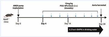
Figure 1: Experimental timeline of AngII-BAPN induced aortic aneurysm formation and PGG-NPs treatment in ApoE-/- mice.
Histological analysis
At the end of the experiment, all surviving mice were sacrificed with inhaled isoflurane before the mice were flushed with 30ml saline. Aortas were exposed with the aid of dissecting microscope to remove periadventitial tissue and photographs were taken. Subsequently, the thoracic and abdominal aorta segments were obtained, fixed in 4% paraformadehyde, and embedded in paraffin wax. Aortas were sectioned with 5uM and stained with hematoxylin and eosin. Elastin assessments were done with aortic sections stained with Verhoeff–Van Gieson (VVG).
Immunofluorescence staining
Paraffin-embedded sections were deparaffinized and rehydrated, followed by permeabilization with 0.1% Triton X-100 (T9284, Sigma-Aldrich, St. Louis, MO, USA) for 10 min. Non-specific binding was blocked using blockBlocker™ solution (NB306, Innovex Biosciences, Richmond, CA, USA). Sections were then incubated overnight at 4°C with primary antibodies against alpha smooth muscle cells (1:200, sc-58671, Santa Cruz Biotechnology, Inc., CA, USA,). For immunofluorescence analysis, sections were incubated with Cy5-conjugated goat anti-rabbit secondary antibodies for 1 h at room temperature, followed by nuclear counterstaining with DAPI (Vector, ZsBio, Beijing, China). Images were acquired using a confocal microscope and quantitative image analysis was performed using ImageJ (NIH, Bethesda, MD, USA).
Statistical analysis
Statistical analyses were performed using R (version 4.2.2). Data are presented as mean ± SEM. Comparisons between two independent groups were performed using an unpaired two-tailed Student’s t-test. Differences in aneurysm incidence were assessed using Fisher’s exact test, and survival curves were compared using the log rank (Mantel–Cox) test. All statistical tests were two-sided, and P < 0.05 was considered statistically significant.
RESULTS
AngII and BAPN induce thoracic and abdominal aortic aneurysm formation and increase mortality in ApoE−/− mice
Combined administration of AngII and BAPN induced aneurysm formation in both the thoracic and abdominal aorta of ApoE−/− mice. Gross examination at day 28 revealed dilation of the thoracic or abdominal aorta in Blank-NP-treated mice, whereas elastin-targeted PGG-NPs treatment markedly attenuated aneurysmal remodeling and maintained gross aortic morphology (Figure 2A). Kaplan–Meier analysis demonstrated progressive mortality in Blank-NP-treated mice throughout the experimental period, while elastin targeted PGG-NPs treatment significantly improved 28-day survival (log rank test, P = 0.0132; Figure 2B). These findings agree with previous reports demonstrating the aggressive nature of the AngII-BAPN model in AAA [11], which is characterized by spontaneous aneurysm rupture and high mortality rates. Quantification of aneurysm incidence further demonstrated the severity of the disease in the Blank NPs-treated group. Incidence analysis showed that AAA developed 75.0% of Blank-NPs-treated mice compared with 23.1% of PGG-treated mice (Fisher’s exact test, P =0.0092). TAA incidence decreased from 25.0% in Blank NPs-treated mice to 15.4% following PGG-NPs treatment, although this difference was not statistically significant (Fisher’s exact test, P = 0.663; Figure 2C).
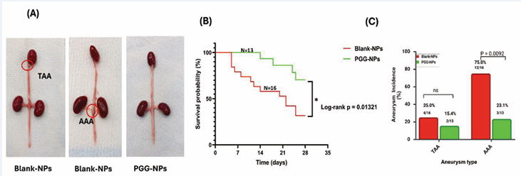
Figure 2: AngII-BAPN induces thoracic and abdominal aortic aneurysms, while PGG nanotherapy improves survival and reduces aneurysm incidence. (A) Representative gross images of aortas harvested from Blank-NP- and PGG-ELN-NP-treated mice following 28 days of AngII-BAPN administration. Blank-NP treated mice exhibited pronounced aneurysmal dilation involving both thoracic (TAA) and abdominal (AAA) aortic segments, whereas PGG-treated mice maintained a more normal aortic morphology. (B)Kaplan–Meier survival analysis of ApoE−/− mice treated with Blank-NPs (n = 16) or PGG-NPs (n = 13) following AngII-BAPN administration. PGG treatment significantly improved 28-day survival compared with Blank-NP-treated controls (log-rank test, P = 0.0132). (C) PGG treatment reduced AAA incidence from 75.0% to 23.1% (P = 0.0092) whereas TAA incidence decreased from 25.0% to 15.4% (P = 0.663). P values were determined using Fisher’s exact test. Data are expressed as the percentage of mice with aneurysms in each treatment group.
AngII-BAPN induces medial remodeling, elastic fiber disruption and vascular smooth muscle cell depletion in thoracic and abdominal aortic aneurysms
Histological analyses revealed marked structural alterations of the aortic wall following AngII-BAPN administration. H&E staining demonstrated pronounced medial remodeling in both thoracic and abdominal aortic segments of Blank-NPs-treated mice, characterized by increased vessel wall thickness (Figure 3A). Quantification of maximal medial thickness confirmed significant increases in both TAA and AAA tissues indicating that AngII-BAPN induces substantial pathological remodeling of the aortic media. In contrast, PGG-NPs-treated mice showed a substantial reduction in wall thickness in TA and AA tissues.
VVG staining further demonstrated extensive disruption of elastic lamellae in Blank-NPs-treated mice. Thoracic and abdominal aortic sections exhibited marked elastin fragmentation and loss of the characteristic concentric organization of the aortic media. In contrast, aortas from PGG-NPs-treated groups maintained a more organized elastic lamellar structure with fewer sites of elastin disruption (Figure 3B). Also, Blank-NPs-treated mice demonstrated reduced αSMA expression in both thoracic and abdominal aneurysms, consistent with VSMC depletion during aneurysm progression. PGG-NPs treatment mitigated the loss of αSMA-positive VSMCs, indicating improved homeostasis and preservation of the contractile phenotype in both thoracic and abdominal aortic segments (Figure 3C).
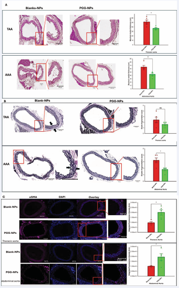
Figure 3: AngII-BAPN induces pathological aortic wall remodeling characterized by medial thickening, elastic fiber disruption and VSMC depletion in thoracic and abdominal aneurysms. (A) H&E staining of thoracic and abdominal aortic sections from Blank-NP- (n=5) and PGG-NP (n=5)-treated mice. Blank-NP treated animals exhibited increased medial thickness, whereas PGG-treated mice demonstrated preservation of a more normal aortic architecture. Quantification of maximal medial thickness demonstrating significant reductions in thoracic and abdominal aortic wall thickness following PGG treatment. Data are presented as mean ± SEM. *P < 0.05. (B) VVG staining of thoracic and abdominal aortas. Elastin fragmentation was scored as: 0, intact; 1, mild; 2, moderate; 3, marked; and 4, severe disruption of elastic lamellae. PGG nanoparticle treatment reduced elastin fragmentation and improved elastic lamellar organization compared with Blank NP-treated mice. (C) Representative αSMA immunofluorescence staining and corresponding fluorescence intensity quantification demonstrating attenuation of contractile VSMC marker loss following PGG treatment in TAA and AAA.
DISCUSSION
Aortic aneurysms have no existing pharmacological therapy [1,2]. Although significant progress has been made in understanding the molecular basis of aneurysm formation, translation of these discoveries into clinically effective treatments has been limited. Most therapeutic approaches investigated to date have focused on suppressing inflammation or inhibiting matrix degradation [13,22-24], with variable success across experimental models but limited translational impact.
This study shows that 10mg/kg elastin-targeted PGG NPs, given systemically, markedly reduced the incidence of thoracic and abdominal aneurysms and significantly improved survival in the AngII-BAPN mouse model. Histological analyses further revealed preservation of elastin, reduced pathological medial remodeling and maintenance of αSMA expression within the aortic media. Earlier studies from our laboratory demonstrated reversal of established elastase-induced AAA following PGG nanoparticle delivery; [9] however, therapeutic efficacy in TAA disease remained unexplored. ECM stabilization observed in both thoracic and abdominal aortic segments, therefore substantially validates our previous findings that PGG nanoparticles interact with monomeric tropoelastin to enhance elastin coacervation, including the activation of vascular SMCs to increase LOX production, both of which are needed for elastic fiber assembly, as the localized presence of PGG facilitates tropoelastin anchoring and LOX crosslinking at the TAA and AAA sites [11,25].
Notably, combined AngII infusion and lysyl oxidase inhibition by BAPN produces one of the most severe forms of experimental aneurysm disease [20,21,26,27]. AngII–BAPN protocols differ in dose, duration and route of administration, producing variable aneurysm distributions and rupture rates. Kanematsu et al., induced thoracic and abdominal aortic aneurysms in male C57BL/6J mice using continuous subcutaneous infusion of AngII at 1,000 ng kg?¹ min?¹ for 6 weeks together with BAPN at 150 mg kg?¹ day?¹ during the first 2 weeks. Both agents were delivered using osmotic minipumps, and combined treatment produced thoracic and abdominal aneurysms with spontaneous rupture, whereas either treatment alone was insufficient to reproduce the full phenotype [20]. Fashandi et al.,subsequently established a rupture-prone model in ApoE deficient mice using AngII at 1,500–2,000 ng kg?¹ min?¹ through subcutaneous osmotic pumps and 0.2% BAPN in drinking water for 28 days, producing high incidences of AAA, TAA and rupture [21]. Jiang et al. used BAPN at 1 g l?¹ in drinking water for 3 weeks before AngII infusion at either 1,000 or 2,500 ng kg?¹ min?¹ and reported greater aortic dissection formation with the higher AngII dose over 14 days [28]. Our regimen of AngII at 1,500 ng kg?¹ min?¹ delivered continuously by subcutaneous osmotic pump together with 0.2%w/v BAPN in drinking water falls within the established rupture-prone range. Untreated mice developed substantial aneurysm burden and progressive mortality throughout the 28-day experimental period. PGG treatment reduced TAA incidence from 25.0% to 15.4% and AAA incidence from 75.0% to 23.1%, suggesting that stabilization of degraded elastin targets pathogenic mechanisms shared throughout the thoracic and abdominal aorta.
Thoracic and abdominal aneurysms differ substantially in developmental origin, ECM composition and underlying molecular mechanisms [29]. Isselbacher proposed that TAA and AAA should be considered distinct disease entities; nevertheless, both ultimately converge on progressive loss of aortic wall integrity [30]. Extensive elastin fragmentation has since emerged as a defining feature of both human and experimental aneurysm disease. Humphrey and colleagues identified elastin degradation as a major determinant of aneurysm progression, demonstrating that disruption of elastic fiber architecture fundamentally alters aortic wall biomechanics and redistributes mechanical stress to remaining medial constituents [31]. As elastic lamellae deteriorates, the surrounding VSMCs experience an increasingly adverse mechanical and biochemical microenvironment, resulting in loss of the contractile phenotype and diminished expression of αSMA, MYH11, and SM22α [32,33]. Reduced VSMC function further impairs extracellular matrix maintenance, thereby establishing a feed-forward axis linking elastin degradation, medial dysfunction and progressive aneurysm expansion. Such a sequence facilitates the concomitant disruption of elastic fibers and reduced αSMA expression observed in untreated mice and suggests that degraded elastin is a common therapeutic substrate throughout the aortic tree. Building upon this paradigm, Isenburg et al. and Parasaram et al., demonstrated that PGG exhibits a high affinity for elastin and limits matrix metalloproteinase-mediated elastic fiber degradation [34,35]. Accordingly, retention of organized elastin together with enhanced αSMA expression following PGG administration is mechanistically aligned with interruption of the pathological cascade linking ECM deterioration and VSMC dysfunction in both elastase and AngII-BAPN models of aortic aneurysm. Demonstration of efficacy in both elastase-induced AAA and AngII BAPN-induced TAA and AAA identifies degraded elastin as a common mechanistic target and positions elastin targeted PGG delivery as a potent ECM-directed strategy for reversing aneurysm progression.
CONCLUSIONS
Aortic aneurysm disease continues to present a substantial clinical burden owing to its often-silent progression and the absence of effective pharmacological interventions. Results from the present study demonstrate that elastin-targeted PGG nanotherapy attenuates the development of thoracic and abdominal aneurysms and confers a significant survival advantage in a rupture prone AngII-BAPN model. Broad efficacy across both elastase- and AngII-BAPN-driven aneurysm paradigms highlights degraded elastin as a convergent node in aneurysm pathobiology and underscores the importance of extracellular matrix homeostasis in determining disease trajectory. Restoration of medial organization and retention of contractile VSMC features further suggest that PGG modulates the interplay between matrix degeneration and vascular remodeling, validating elastin-directed nanomedicine as a compelling avenue for aneurysm intervention and laying the foundation for advancing matrix-centric therapies toward clinical translation.
Funding: This work was funded by NIH (HL133662) to N.R.Vyavahare.
Conflict of interest: N. R. Vyavahare is a major shareholder in Elastrin Therapeutics Inc., a company that has licensed the nanoparticle therapy from Clemson University.
Acknowledgments: The authors thank Agnes Nagy Mehesz and Guzeliya Korneva for their valuable support and assistance throughout the development of this work.
REFERENCES
- Ma G, Li Y, Jia F. The global burden of aortic aneurysm in adults over 55: evolving trends, risk factors, and projections. Front Cardiovasc Med. 2025; 12: 1629635.
- Wu H, Xie C, Wang R, Cheng J, Xu Q, Zhao H. Comparative analysis of thoracic and abdominal aortic aneurysms across the segment and species at the single-cell level. Front Pharmacol. 2023; 13: 1095757.
- Scali ST, Stone DH. Modern management of ruptured abdominal aortic aneurysm. Front Cardiovasc Med. 2023; 10: 1323465.
- Atkinson G, Bianco R, Di Gregoli K, Johnson JL. The contribution of matrix metalloproteinases and their inhibitors to the development, progression, and rupture of abdominal aortic aneurysms. Front Cardiovasc Med. 2023; 10: 1248561.
- Cai D, Chen SY. Elastin in the Pathogenesis of Abdominal Aortic Aneurysm. Cells. 2025; 14: 1597.
- Harky A, Fan KS, Fan KH. The genetics and biomechanics of thoracic aortic diseases. Vasc Biol. 2019; 1: R13-R25.
- Crandall CL, Caballero B, Viso ME, Vyavahare NR, Wagenseil JE. Pentagalloyl Glucose (PGG) Prevents and Restores Mechanical Changes Caused by Elastic Fiber Fragmentation in the Mouse Ascending Aorta. Ann Biomed Eng. 2023; 51: 806-819.
- Pavey SN, Cocciolone AJ, Marty AG, Ismail HN, Hawes JZ, Wagenseil JE. Pentagalloyl glucose (PGG) partially prevents arterial mechanical changes due to elastin degradation. Exp Mech. 2021; 61: 41-51.
- Dhital S, Rice CD, Vyavahare NR. Reversal of elastase-induced abdominal aortic aneurysm following the delivery of nanoparticle-based pentagalloyl glucose (PGG) is associated with reduced inflammatory and immune markers. Eur J Pharmacol. 2021; 910: 174487.
- Bhamidipati CM, Mehta GS, Lu G, Moehle CW, Barbery C, DiMusto PD, et al. Development of a novel murine model of aortic aneurysms using peri-adventitial elastase. Surgery. 2012; 152: 238-246.
- Wang X, Parasaram V, Dhital S, Nosoudi N, Hasanain S, Lane BA, et al. Systemic delivery of targeted nanotherapeutic reverses angiotensin II-induced abdominal aortic aneurysms in mice. Sci Rep. 2021; 11: 8584.
- Ren W, Liu Y, Wang X, Jia L, Piao C, Lan F, et al. β-Aminopropionitrile monofumarate induces thoracic aortic dissection in C57BL/6 mice. Sci Rep. 2016; 6: 28149.
- Yin L, Kent EW, Wang B. Progress in murine models of ruptured abdominal aortic aneurysm. Front Cardiovasc Med. 2022; 9: 950018.
- Kawai T, Takayanagi T, Forrester SJ, Preston KJ, Obama T, Tsuji T, et al. Vascular ADAM17 (a Disintegrin and Metalloproteinase Domain 17) Is Required for Angiotensin II/β-Aminopropionitrile-Induced Abdominal Aortic Aneurysm. Hypertension. 2017; 70: 959-963.
- Ren W, Liu Y, Wang X, Jia L, Piao C, Lan F, et al. β-Aminopropionitrile monofumarate induces thoracic aortic dissection in C57BL/6 mice. Sci Rep. 2016; 6: 28149.
- Ortega R, Collado A, Selles F, Gonzalez-Navarro H, Sanz MJ, Real JT, et al. SGLT-2 (Sodium-Glucose Cotransporter 2) Inhibition Reduces Ang II (Angiotensin II)-Induced Dissecting Abdominal Aortic Aneurysm in ApoE (Apolipoprotein E) Knockout Mice. Arterioscler Thromb Vasc Biol. 2019; 39: 1614-1628.
- Sawada H, Lu HS, Cassis LA, Daugherty A. Twenty Years of Studying AngII (Angiotensin II)-Induced Abdominal Aortic Pathologies in Mice: Continuing Questions and Challenges to Provide Insight Into the Human Disease. Arterioscler Thromb Vasc Biol. 2022; 42: 277-288.
- Cassis LA, Gupte M, Thayer S, Zhang X, Charnigo R, Howatt DA, et al. ANG II infusion promotes abdominal aortic aneurysms independent of increased blood pressure in hypercholesterolemic mice. Am J Physiol Heart Circ Physiol. 2009; 296: H1660-H1665.
- Fashandi AZ, Spinosa M, Salmon M, Su G, Montgomery W, Mast A, et al. Female Mice Exhibit Abdominal Aortic Aneurysm Protection in an Established Rupture Model. J Surg Res. 2020; 247: 387-396.
- Kanematsu Y, Kanematsu M, Kurihara C, Tsou TL, Nuki Y, Liang EI, et al. Pharmacologically induced thoracic and abdominal aortic aneurysms in mice. Hypertension. 2010; 55: 1267-1274.
- Fashandi AZ, Hawkins RB, Salmon MD, Spinosa MD, Montgomery WG, Cullen JM, et al. A novel reproducible model of aortic aneurysm rupture. Surgery. 2018; 163: 397-403.
- Kavaliunaite E, Sejr Skovbo Kristensen J, Scheurer S, Berg I, Lindholt JS, Stubbe J. Therapeutic Strategies for Abdominal Aortic Aneurysm: A Comprehensive Systematic Review. J Cardiovasc Dev Dis. 2025; 12: 462.
- Shaikh II, Singh S, Feng Y, Shahzad KA, Wang J, Zhou Q, et al. Therapeutic Targeting of Signaling Pathways in Abdominal Aortic Aneurysm: From Pathogenesis to Precision Medicine. Drug Des Devel Ther. 2025; 19: 12077-12111.
- Cameron SJ, Russell HM, Owens AP. Antithrombotic Therapy in Abdominal Aortic Aneurysm: Beneficial or Detrimental? 2018.
- Dhital S, Vyavahare NR. Nanoparticle-based targeted delivery of pentagalloyl glucose reverses elastase-induced abdominal aortic aneurysm and restores aorta to the healthy state in mice. PLoS One. 2020; 15: e0227165.
- Sawada H, Beckner ZA, Ito S, Daugherty A, Lu HS. β-Aminopropionitrile-induced aortic aneurysm and dissection in mice. JVS-Vascu Sci. 2022; 3: 64-72.
- Hao X, Wu S, Cheng S, Wang X, Zhang J, Li X, et al. Mouse models of aortic dissection induced by β-aminopropionitrile combined with angiotensin II and the changes in their gut microbiota. BMC Microbiol. 2025; 25: 419.
- Jiang H, Liu W, He X, Zeng H. Establishment of a meta-analysis based novel aortic dissection mouse model. Sci Rep. 2022; 12: 21434.
- Gyftopoulos A, Ziganshin BA, Elefteriades JA, Ochoa Chaar CI. Comparison of Genes Associated with Thoracic and Abdominal Aortic Aneurysms. Aorta (Stamford). 2023; 11: 125-134.
- Isselbacher EM. Thoracic and abdominal aortic aneurysms. Circulation. 2005; 111: 816-828.
- Humphrey JD, Holzapfel GA. Mechanics, mechanobiology, and modeling of human abdominal aorta and aneurysms. J Biomech. 2012; 45: 805-814.
- Cao G, Xuan X, Hu J, Zhang R, Jin H, Dong H. How vascular smooth muscle cell phenotype switching contributes to vascular disease. Cell Commun Signal. 2022; 20: 180.
- Pai A, Leaf EM, El-Abbadi M, Giachelli CM. Elastin degradation and vascular smooth muscle cell phenotype change precede cell loss and arterial medial calcification in a uremic mouse model of chronic kidney disease. Am J Pathol. 2011; 178: 764-773.
- Parasaram V, Nosoudi N, Chowdhury A, Vyavahare N. Pentagalloyl glucose increases elastin deposition, decreases reactive oxygen species and matrix metalloproteinase activity in pulmonary fibroblasts under inflammatory conditions. Biochem Biophys Res Commun. 2018; 499: 24-29.
- Isenburg JC, Simionescu DT, Starcher BC, Vyavahare NR. Elastin stabilization for treatment of abdominal aortic aneurysms. Circulation. 2007; 115: 1729-1737.