Early-Onset Carcinoid Heart Disease: A Rare Presentation in Youth
- 1. College of Medicine, University of Tennessee Health Science Center, USA
- 2. Department of Internal Medicine, University of Tennessee Health Science Center
Abstract
Neuroendocrine tumors (NETs) may present in various ways, including as a result of carcinoid syndrome (CS). Typical manifestations of CS include chronic flushing and/or diarrhea, though a well-established complication is carcinoid heart disease (CHD) which has a profound impact on morbidity and mortality. We present a 21-year-old male hospitalized for acute onset of decompensated right-sided heart failure (RHF) due to underlying CS. Our case emphasizes the importance of considering CS in the differential diagnosis for patients presenting with right-sided heart failure, particularly in the absence of common risk factors such as advanced age and ischemic heart disease.
Citation
Elliot J, Ammon J, Bejnood A, Coleman A, Dunlap N (2026) Early-Onset Carcinoid Heart Disease: A Rare Presentation in Youth. J Cardiol Clin Res. 14(2): 1224.
BACKGROUND
Carcinoid syndrome (CS), a paraneoplastic syndrome associated with hormonally active neuroendocrine tumors (NETs), manifests in 30-40% of NET patients [1]. Most primary tumors originate in the gastrointestinal (GI) tract, releasing hormones such as serotonin, which exert local effects prior to hepatic metabolism to 5-hydroxyindoleacetic acid (5-HIAA) with subsequent urinary excretion [2].
As tumors metastasize to the liver, circumventing metabolism, systemic effects emerge. These often involve the heart, notably affecting right-sided heart valves contributing to carcinoid heart disease (CHD). Other manifestations include bronchospasm, dyspnea, and episodic cutaneous flushing. While vasoactive hormones are metabolized in the lungs, typically sparing the left sided heart valves, their effects on right-sided heart valves cause deposition of fibrous tissue on valves, papillary muscles, and ventricular walls, precipitating dysfunction [3]. Severe tricuspid valve damage may present as acute onset of decompensated right-sided heart failure (RHF), as seen in this case.
The mean age of patients with GI carcinoid tumors is 60, with CHD typically affecting individuals between 50 and 70-years-old [3]. Despite an escalating incidence rate outpacing other cancer types [4], prognosis has improved, leading to higher incidence of long-term complications such as CHD [5]. CHD is diagnosed in 50% of CS patients, with approximately 20% initially presenting with CHD [6]. Unfortunately, CHD is frequently diagnosed late due to inadequate screening strategies, often after symptomatic onset [7].This case serves to illustrate how difficult it can be to arrive at this already rare diagnosis in a patient that does not fit the usual demographic profile.
OBJECTIVE
Our goal is to contribute to the limited literature of CS in young patients (especially ≤ 21-years-old), with hopes of increasing clinician suspicion for CS when generating a differential diagnosis in young patients with RHF.
CASE REPORT
A 21-year-old Spanish speaking male with no known past medical history presented to the emergency department with progressive dyspnea and lower extremity edema that began 4 months prior. He had recently migrated from a Central American country and had limited access to medical care.
On arrival, vital signs were remarkable for heart rate of 120 beats/min and respiratory rate of >20 breaths/ min. Oxygen saturation (SpO2) remained >95%, however he appeared uncomfortable. Patient noted to have jugular venous distention to the level of the mandible with a positive hepatojugular reflux; he had a harsh, grade III/ VI systolic murmur best heard at the right lower sternal border with radiation to all other areas of auscultation. Lung auscultation was clear bilaterally. He had pitting edema of bilateral lower extremities extending to his hip. A prominent erythematous rash was present, primarily involving the face, neck, chest, and upper extremities. Review of systems was positive for persistent diarrhea of 4 years.
Routine blood tests and cardiac biomarkers were obtained; findings included alkaline phosphatase of 226 IU/L, aspartate aminotransferase of 26 IU/L, alanine aminotransferase of 39 IU/L, B-type natriuretic peptide of 173 pg/mL, and a high-sensitivity troponin of 4 pg/mL. Thyroid stimulating hormone level was 2.34 mIU/L.
Electrocardiogram showed sinus tachycardia, right axis deviation, and incomplete right bundle branch block. The chest radiograph did not show any abnormal findings. Transthoracic echocardiogram (TTE) demonstrated a left ventricular ejection fraction of 55-60% and severe tricuspid regurgitation (Figure 1).
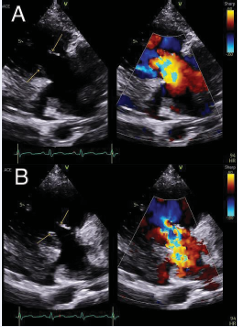
Figure 1: Tricuspid valve leaflets (yellow arrows) during both cardiac diastole (A) and systole (B). There is poor coaptation of the tricuspid leaflets, as well as significant tricuspid regurgitation seen on color doppler.
A computed tomography angiography was obtained and negative for pulmonary embolism, though incidentally it showed multiple low attenuation hepatic masses. Magnetic resonance imaging (MRI) of the abdomen revealed diffuse hepatic metastatic disease with mesenteric lymphadenopathy as well as right posterior hepatic vein occlusion secondary to mass effect (Figure 2).
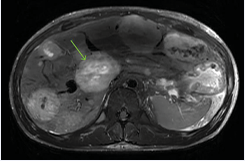
Figure 2: MRI of abdomen and pelvis with and without contrast seen in axial plane T2 weighted demonstrating an enlarged liver with numerous scattered hepatic lesions, concerning for malignancy. The largest lesion is highlighted by the green arrow.
The patient’s initial clinical presentation in the setting of severe tricuspid regurgitation and multiple hepatic lesions was suggestive of CHD secondary to a GI primary NET.
An initial diagnostic test of 24-hour urinary excretion of 5-HIAA was collected and significantly elevated at 444.3 mg/24 hours. A subsequent liver biopsy demonstrated a well-differentiated neuroendocrine tumor, grade 1. Various tumor markers were tested; they were positive for CK8/18, synaptophysin, chromogranin, INSM-1, and negative for HepPar1 and CD34.
The goal of initial treatment was symptom management. The dyspnea and lower extremity edema improved with intravenous diuretics, and he was ultimately transitioned to oral diuretics to maintain euvolemia. Octreotide was initiated with subsequent improvement of his cutaneous flushing and diarrhea. Given his clinical improvement, he was discharged on these medications with arrangements to follow-up with oncology outpatient and discuss chemotherapy. Additionally, he was referred to cardiothoracic surgery for surgical tricuspid valve replacement.
DISCUSSION
While CS and CHD are rare, both are well described in literature. CS remains a very uncommon diagnosis; there is an estimated prevalence of 1.2 to 2.1 cases of carcinoid tumors per 100,000 people, and of these only 20-30% present with CS. Of the CS cases, about half present with CHD [8]. In other words, between 1.2 and 3.2 cases of CHD exist per 1,000,000 people. They are most frequently diagnosed in ages 50-69, however it is also estimated that the average time to diagnosis is at least 9 years – diagnosis typically happens after metastasis to the liver [9].
As mentioned, the disease process typically affects an older population, and many patients with cardiac involvement typically demonstrate a slow progression of symptoms [10].This patient demonstrated prominent features of heart failure upon presentation, which in young individuals often prompts consideration of more common etiologies such as congenital heart disease or viral myocarditis. Therefore, the association between carcinoid syndrome and heart failure may not be immediately apparent, leading to potential delays in diagnosis. Subsequently, clinicians should consider the possibility of carcinoid syndrome in young patients who present with new heart failure with at least one other symptom potentially indicative of carcinoid (flushing, diarrhea, bronchospasm, nausea/vomiting). As part of this evaluation, it may be reasonable to perform a computerized tomography (CT) scan of the abdomen/pelvis to evaluate for possible tumors. Additionally, a 24 hour urine 5-HIAA study may be warranted if clinical suspicion is high. Lastly, it may be reasonable to perform TTE/TEE in patients diagnosed with (or who have suspicion for having) carcinoid tumors to screen for CHD.
This patient’s symptoms progressed for months prior to seeking medical attention, and many psychosocial factors may have contributed to his delay in presentation. Prior surveys of Latino(a)/Latinx community members have cited reasons for delay in seeking medical care. These include generalized fear resulting from anti-immigrant rhetoric, behavior change due to threat of immigration enforcement, financial and opportunity costs related to healthcare access, and culturally discordant health seeking practices by country of origin [11]. Clinicians should be cognizant of the psychosocial aspects that may result in delayed disease presentations in Latino(a)/Latinx populations.
In conclusion, the case of this young patient with carcinoid heart disease underscores the challenges that can arise in diagnosing CHD. The medical team faced multiple obstacles in uncovering this patient’s diagnosis, including an uncommon presentation, prominent psychosocial factors, and healthcare access disparities that hindered care in a vulnerable patient population. Clinicians must be diligent in maintaining a broad differential so as not to miss an uncommon presentation of a rare disease.
ACKNOWLEDGMENTS
James Elliot, Jessica Ammon, Aram Bejnood, Adam Coleman, and Natalie Dunlap prepared the manuscript.
Natalie Dunlap finalized the manuscript.
All authors approved and endorsed the content of this manuscript.
REFERENCES
- Rubin de Celis Ferrari AC, Glasberg J, Riechelmann RP. Carcinoid syndrome: update on the pathophysiology and treatment. Clinics (Sao Paulo). 2018; 73: e490s.
- Srirajaskanthan R, Shanmugabavan D, Ramage JK. Carcinoid syndrome. BMJ. 2010; 341: c3941.
- Jin C, Sharma AN, Thevakumar B, Majid M, Takahashi N, Tanious A, et al. Carcinoid Heart Disease: Pathophysiology, Pathology, Clinical Manifestations, and Management. Cardiology. 2021; 146: 65-73.
- Mocellin S, Nitti D. Gastrointestinal carcinoid: epidemiological and survival evidence from a large population-based study (n = 25 531). Ann Oncol. 2013; 24: 3040-3044.
- Zuetenhorst JM, Taal BG. Metastatic carcinoid tumors: a clinical review. Oncologist. 2005; 10: 123-131.
- Hassan SA, Banchs J, Iliescu C, Dasari A, Yusuf SW. Carcinoid heart disease. Heart. 2017; 103: 1488-1495.
- Laskaratos FM, Davar J, Toumpanakis C. Carcinoid Heart Disease: a Review. Curr Oncol Rep. 2021; 23: 48.
- Matshela MR. Cases in a series of carcinoid syndrome and carcinoid heart disease. Cardiovasc J Afr. 2018; 29: e1-e7.
- Horton KM, Kamel I, Hofmann L, Fishmann EK. Carcinoid tumors of the small bowel: a multitechnique imaging approach. AJR Am J Roentgenol. 2004; 182: 559-567.
- Mota JM, Sousa LG, Riechelmann RP. Complications from carcinoid syndrome: review of the current evidence. Ecancermedicalscience. 2016; 10: 662.
- Doshi M, Lopez WD, Mesa H, Bryce R. Barriers & facilitators to healthcare and social services among undocumented Latino(a)/ Latinx immigrant clients: Perspectives from frontline service providers in Southeast Michigan. PLoS One. 2020; 15: e0233839.