Intravascular Lithotripsy as Salvage Therapy for Heavily Calcified Carotid Artery Stenosis during Carotid Artery Stenting: A Case Report
- 1. Department of Neurosurgery, Mass General Brigham, Harvard Medical School, USA
- 2. Virgin Research Institute, México
- 3. Facultad de Medicina, Universidad Autonoma de Guadalajara, Mexico
- 4. Beth Israel Deaconess Medical Center, Harvard Medical School, USA
Abstract
Stroke remains a leading cause of death and disability worldwide. In appropriately selected patients with symptomatic carotid artery stenosis, carotid artery stenting (CAS) is an established strategy for stroke prevention. However, severe vascular calcification may limit adequate lesion preparation, resulting in suboptimal stent expansion, thereby compromising procedural success. Although intravascular lithotripsy (IVL) has emerged as a plaque-modification technique for heavily calcified vascular lesions, clinical experience in the carotid circulation remains limited. We report the case of an 83-year-old woman with symptomatic critical right internal carotid artery stenosis who underwent CAS. After unsuccessful lesion preparation with cutting balloon angioplasty, IVL was used as salvage therapy before stent deployment. IVL facilitated adequate plaque modification, allowing stent expansion and a satisfactory angiographic result following adjunctive post-dilation. The only procedural complication was transient bradycardia, which resolved promptly with atropine. The patient experienced no perioperative neurological complications and remained free of recurrent stroke during 12 months of follow-up. This case illustrates the potential role of IVL as a salvage strategy when conventional lesion preparation during CAS is unsuccessful and supports its consideration in selected patients with heavily calcified carotid lesions.
Keywords
• Carotid artery stenosis
• Intravascular lithotripsy
• Carotid artery stenting
• Vascular calcification
• Salvage therapy
• Stroke
Citation
Hernandez DA, Avila R, Zuloaga A, Salido JG, Pacheco N, et al. (2026) Intravascular Lithotripsy as Salvage Therapy for Heavily Calcified Carotid Artery Stenosis during Carotid Artery Stenting: A Case Report. J Neurol Disord Stroke 13(2): 1251.
ABBREVIATIONS
Fr: French; IVL: Intravascular Lithotripsy
INTRODUCTION
Stroke is one of the leading causes of death and disability worldwide [1]. From the age of 25 years, approximately one in four individuals will experience a stroke during their lifetime [2]. Each year, an estimated 11.9 million people suffer a new stroke, and approximately 7.3 million die from the disease [1]. Stroke can be classified into several subtypes, the most common of which is ischemic stroke. Carotid artery disease (CAD) accounts for a substantial proportion of ischemic strokes, causing approximately 8–15% of all cases [3].The current standard of care for CAD consists of optimal medical therapy and, in selected patients, carotid revascularization. Appropriate revascularization can significantly improve outcomes and reduce the risk of recurrent stroke [4].
Endovascular approaches are well established as part of the standard of care; however, despite substantial advances in endovascular techniques and device technology, severe arterial calcification remains one of the greatest technical challenges in carotid artery stenting (CAS). Extensive circumferential calcification reduces arterial compliance, impairs adequate lesion preparation, and limits stent expansion, ultimately leading to suboptimal procedural outcomes and carotid artery stenting failure [5,6]. Management of heavily calcified carotid lesions remains challenging, and available plaque-modification techniques are limited, with cutting and scoring balloons representing the most commonly reported endovascular strategies before stent deployment [4,7]. Intravascular lithotripsy (IVL) has emerged as a novel plaque-modification technology capable of delivering pulsatile acoustic pressure waves that selectively fracture superficial and deep vascular calcium while minimizing injury to surrounding soft tissue [8,9]. Currently, IVL is approved by the U.S. Food and Drug Administration (FDA) for coronary and peripheral arterial disease, whereas evidence supporting its application in carotid artery disease remains limited to observational studies and case series [9,10]. Here, we present our experience using IVL as a salvage therapy following failed cutting balloon angioplasty in a patient with severe calcific carotid artery disease and marked internal carotid artery (ICA) tortuosity, with particular emphasis on the technical considerations required for the safe and effective use of this technology.
CASE REPORT
An 83-year-old woman with a past medical history of type two diabetes mellitus, hypertension and hypercholesterolemia, managed with insulin, dual antiplatelet therapy and atorvastatin, presented with acute left-sided facial drop, blurred vision, left upper extremity weakness 2/5, left lower extremity weakness 3/5 accompanied with hemisensory loss and pronator drift. Computed tomography angiography (CTA) demonstrated a right sided middle cerebral artery stroke and a 95% stenosis at the proximal segment of the right ICA with extensive circumferential calcification measuring 4 mm in thickness (Figure 1), as well as marked tortuosity of the distal cervical internal carotid artery (Figure 2).
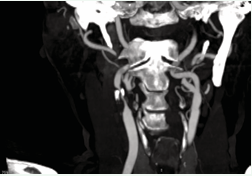
Figure 1 Coronal computed tomography angiography (CTA) demonstrating critical stenosis of the right internal carotid artery at the carotid bifurcation (arrow), with extensive circumferential calcification.
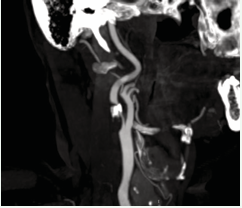
Figure 2: Sagittal CTA showing marked tortuosity of the cervical right internal carotid artery, an anatomical feature associated with increased technical complexity during carotid artery stenting.
After performing medical reperfusion therapy for acute stroke management and five days after symptom onset digital subtraction angiography demonstrated preserved intracranial perfusion and confirmed the same findings encountered in the CTA. Based on the clinical and imaging findings, the diagnosis of critical right internal carotid artery stenosis was established. A multidisciplinary team reached a consensus not to pursue CEA due to the high risk features of the patient and following standard procedure, carotid artery stenting was performed under local anesthesia via ultrasound-guided access to the right common femoral artery. A 5-Fr Judkins Right (JR) 3.5 catheter was advanced over a 0.035-inch hydrophilic guidewire to selectively catheterize the right common carotid artery. The guidewire was exchanged for a 0.035 inch Amplatz stiff guidewire, allowing advancement of a long 7-Fr introducer sheath into the common carotid artery. Systemic anticoagulation was achieved with 6,000 units of intravenous unfractionated heparin. Following diagnostic angiography, a Filter EZ distal embolic protection device was deployed in the distal internal carotid artery beyond the target lesion to provide cerebral embolic protection during intervention. Initial lesion preparation was attempted using a 2.5 × 10 mm cutting balloon inflated to its nominal pressure of 8 atm. Despite cutting balloon angioplasty, satisfactory lesion expansion was not achieved. The lesion was therefore treated with intravascular lithotripsy (IVL) as a salvage strategy to further modify the lesion before definitive stent implantation. A 4 × 40 mm Shockwave IVL balloon was selected using a 1:1 balloon-to-vessel sizing strategy to optimize calcium modification while minimizing injury to the adjacent normal carotid artery, delivering 30 shockwave pulses (Figure 3).
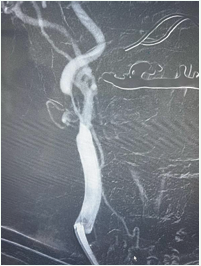
Figure 3: Intraoperative angiography demonstrating positioning of the intravascular lithotripsy balloon across the calcified right internal carotid artery stenosis before energy delivery. During lithotripsy, the patient developed transient bradycardia, which resolved promptly after administration of 1 mg of intravenous atropine.
During IVL, the patient developed transient bradycardia, which resolved promptly following administration of 1 mg of intravenous atropine. Following successful plaque modification, a 7–10 × 40 mm self-expanding carotid stent was deployed across the stenotic segment. Completion angiography demonstrated approximately 40% residual stenosis, prompting additional post-dilation with a 5 × 20 mm angioplasty balloon inflated to 12 atm. Final angiography demonstrated satisfactory stent expansion with adequate luminal gain and <30% residual stenosis (Figure 4). The patient experienced transient bradycardia during the first 24 hours after the procedure but remained neurologically stable without perioperative stroke or other procedural complications and was discharged the following day on dual antiplatelet therapy and continued to do well without recurrent stroke at 3, 6 and 12-month follow-up.
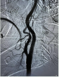
Figure 4: Final angiogram demonstrating satisfactory expansion of the carotid stent with restoration of luminal patency.
DISCUSSION
In selected patients who are poor candidates for both carotid endarterectomy and conventional carotid artery stenting (CAS), intravascular lithotripsy (IVL) represents a promising plaque-modification therapy. Although IVL is currently approved only for coronary and peripheral arterial disease, its application in the carotid circulation remains off-label, and the available evidence is limited to observational studies [9]. Several technical considerations should be highlighted when performing carotid IVL.
Unlike conventional balloon angioplasty, IVL requires prolonged balloon inflations of approximately 30 seconds to deliver one lithotripsy pulse per second, often necessitating multiple treatment cycles [8]. Consequently, prolonged carotid sinus stimulation may provoke vagally mediated bradycardia with transient hemodynamic instability, as observed in our patient and previously reported in the literature [10,11]. Appropriate balloon sizing is equally important. A 1:1 IVL balloon-to-reference vessel diameter ratio is recommended to maximize plaque modification while minimizing vessel injury [8]. However, the currently available Shockwave S4 catheter is limited to a maximum balloon diameter of 4.0 mm, whereas the normal internal carotid artery typically measures 4.5–6.0 mm in diameter [12]. In our case, this limitation resulted in residual stenosis requiring post-dilation with a 5 × 20 mm angioplasty balloon, highlighting the need for a carotid specific IVL platform with larger balloon diameters.
Conversely, the larger Shockwave M5 catheter offers balloon diameters of up to 7.0 mm but has a shorter working length and a larger crossing profile, which may limit its deliverability in tortuous anatomy or during transfemoral access [8]. Furthermore, because IVL catheters have a larger crossing profile than conventional angioplasty balloons, predilation with a smaller balloon is often necessary to facilitate device delivery [8]. The characteristics of the currently available Shockwave IVL catheter platforms are summarized in Table 1.
Table 1: Comparison of currently available Shockwave intravascular lithotripsy (IVL) catheter platforms.
|
Characteristic |
Shockwave C2 |
Shockwave S4 |
Shockwave M5 |
|
|
(Coronary) |
(Small Peripheral) |
(Medium Peripheral) |
|
Balloon diameter |
2.5–4.0 mm |
2.5–4.0 mm |
3.5–7.0 mm |
|
Balloon length |
12 mm |
40 mm |
60 mm |
|
Crossing profile |
0.044–0.047 in |
0.048–0.050 in |
0.054–0.073 in |
|
Sheath compatibility |
6 Fr guide catheter |
5 Fr |
6 Fr (3.5–6.0 mm) |
|
|
|
|
7 Fr (6.5–7.0 mm) |
|
Working length |
138 cm |
135 cm |
110 cm |
|
Guidewire compatibility |
0.014 in |
0.014 in |
0.014 in |
|
Lithotripsy emitters |
1 |
2 |
3 |
|
Maximum pulses per catheter |
80 |
160 |
300 |
|
Recommended balloon sizing |
1:1 to reference vessel diameter |
1.1:1 to reference vessel diameter |
1.1:1 to reference vessel diameter |
Although IVL appears to be a valuable adjunct for the treatment of heavily calcified carotid lesions, the therapy is resource-intensive, and important questions remain regarding its long-term durability, the optimal lithotripsy pulse regimen, patient selection, and its comparative effectiveness relative to other plaque-modification strategies [9]. Prospective multicenter studies are needed to better define the role of IVL in carotid revascularization and facilitate the development of carotid-specific IVL devices.
CONCLUSION
This case illustrates the potential role of intravascular lithotripsy (IVL) as a salvage strategy when conventional lesion preparation during carotid artery stenting (CAS) is unsuccessful. By facilitating adequate plaque modification after failed cutting balloon angioplasty, IVL enabled successful stent deployment without perioperative neurological complications. Although experience with IVL in the carotid circulation remains limited, this case supports its consideration as a salvage option in selected patients with heavily calcified carotid lesions.
ACKNOWLEDGEMENTS
The authors wish to thank Virgin Research Institute involved in the care of the patient and the department of Cardiology of Hospital Angeles del Carmen for providing the diagnostic images. We also acknowledge our medical school faculties for providing us with the resources needed to conduct the literature review needed to write this manuscript.
Ethical Approval
Ethical approval was not required for this case report in accordance with the policies of Virgin Research Institute.
Informed Consent: Written informed consent was obtained from the patient.
Data Accessibility Statement: Data sharing is not applicable to this case report, since the only available data for this manuscript is the medical record, which is confidential.
REFERENCES
- GBD 2021 Stroke Risk Factor Collaborators. Global, regional, and national burden of stroke and its risk factors, 1990–2021: a systematic analysis for the Global Burden of Disease Study 2021. Lancet Neurol. 2024; 23: 973-1003.
- GBD 2016 Lifetime Risk of Stroke Collaborators, Feigin VL, Nguyen G. Global, regional, and country-specific lifetime risks of stroke, 1990 and 2016. N Engl J Med. 2018; 379: 2429-2437.
- Bonati LH, Jansen O, de Borst GJ, Brown MM. Management of atherosclerotic extracranial carotid artery stenosis. Lancet Neurol. 2022; 21: 273-283.
- AbuRahma AF, Avgerinos ED, Chang RW. Society for Vascular Surgery clinical practice guidelines for management of extracranial cerebrovascular disease. J Vasc Surg. 2022; 75: 26S-98S.
- Elsayed N, Yei KS, Naazie I. The impact of carotid lesion calcification on outcomes of carotid artery stenting. J Vasc Surg. 2022; 75: 897-904.
- DiLosa K, Schonefeld S, El-Khoury R. Multi-center experience with intravascular lithotripsy for treatment of severe calcification during transcarotid artery revascularization for high-risk patients. J Vasc Surg. 2024; 80: 815-822.
- Castriota F, de Campos Martins EC, Setacci C. Cutting balloon angioplasty in percutaneous carotid interventions. J Endovasc Ther. 2008; 15: 621-631.
- Kereiakes DJ, Virmani R, Hokama JY. Principles of intravascular lithotripsy for calcific plaque modification. JACC Cardiovasc Interv. 2021; 14: 1275-1292.
- Hernandez Langarica DA, Gonzalez-Salido J, Hernandez AJ, Kappel A, Virgen LR. Intravascular lithotripsy in calcified carotid stenosis: technical and clinical outcomes. J Neurointerv Surg. 2026; 18: 1347-1354.
- Mitchell M, Verhey LH, Taylor N. Intravascular lithotripsy followed by angioplasty and stenting for severely calcified carotid artery stenosis: technical note and case series. Front Neurol. 2025; 16: 1573413.
- Hanel RA, Jaikumar V, Gutierrez-Aguirre SF, et al. Adjunctive intravascular lithotripsy for heavily calcified carotid stenosis: a dual-center experience and technical case series. J Neurointerv Surg. 2025; 17: e42.
- Krejza J, Arkuszewski M, Kasner SE. Carotid artery diameter in men and women and the relation to body and neck size. Stroke. 2006; 37: 1103-1105.











































































































































