Early Return of Vision after the Use of Oral Corticosteroids in Macular Burns by Looking at the Sun
- 1. Department of Ophthalmology, Birjand University of Medical Science, Iran
Abstract
A report of early vision return in a patient who had a macular burn after looking at the sun and was prescribed oral steroids. In the ophthalmological examination, the uncorrected visual acuity of both eyes was 10/10, in the fundus examination, a yellowish white lesion was evident in the center of the macula of both eyes, and a hyperreflective lesion was observed in the OCT. After three days of treatment, the patient’s visual acuity improved significantly. Although there was no change in the fundus examination, the fundus imaging and OCT that were performed two weeks later also improved.
Keywords
• Solar maculopathy
• Causes
• Treatment
CITATION
Gholamhossein Y (2024) Early Return of Vision after the Use of Oral Corticosteroids in Macular Burns by Looking at the Sun. JSM Ophthalmol 11(1): 1091.
CASE PRESENTATION
A 25-year-old man who had no eye disease in his history and had visual impairment in both eyes after looking at the sun for a quarter of an hour, went to the ophthalmology clinic and during the examination, each eye had an acuity of 6.10. They had vision, and in the fundus examination, a yellowish white spot was evident in the center of both maculae. Also, OCT showed hyperreflectivity in the outer part of the retina of both eyes. Two weeks after the onset of this complication, the visual acuity of each eye has improved significantly, and the vision is between 9.10 and 10.10. However, in ophthalmoscopy, the lesion is improving, and in OCT, the amount of hyperreflectivity has decreased. There are not few similar reports that despite repeated recommendations, this happens for various and preventable reasons, such as looking at the solar eclipse, rainbow or laser pointer or looking at the sun [1] (Figure 1).
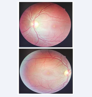
Figure 1: Fundus photo of right and left eye showed yellow spot in center of macula.
In one report, a one-sided case was reported by Taylor, although this event can cause both eyes to be identical, as in our case, or non-identical, as in Taylor’s report [2], that it is not possible to justify the conflict in one case, and several reasons may play a role in it, including the state of refractive errors or disturbances in the transparent environment of the eye, or causes that are not known, play a role in it, the findings of oct in this case The damage was in the part below the retina In a study conducted in India, the finding of oct was also similar to the same report [3] (Figure 2).
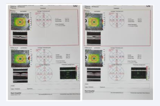
Figure 2: Retina OCT of right and left eye showed early hyperreflectivity and one week thereafter hyporeflectivity spot in center of macula in outer retina
In another study, the improvement of vision depends on the early improvement of the external limiting membrane (ELM) in the area before the oval area and the interstitial area of the outer parts of the retina which inferences in this study have similar results in other studies related to the integrity of the ELM in improving visual acuity in Other macular diseases are related [4].
And in the case of our report, the transparent environment in the eye based on the red reflex in both eyes was normal and the same, and the refraction in both eyes had no refractive error. Early vision return was also associated with improvement in the external limiting membrane. Currently, our one case report may bring promise in evaluating the therapeutic effects of oral corticosteroids and early vision return.


