Gentisic Acid Enhances Porcine Oocyte Maturation Quality and Developmental Competence of Somatic Cell Nuclear Transfer by Decreasing Oxidative Stress
- 1. Faculty of Biotechnology, Jeju National University, Republic of Korea
- 2. Stem Cell Research Center, Jeju National University, Republic of Korea
- 3. Subtropical Livestock Research Institute, National Institute of Animal Science, Korea
- 4. Bio Medical, College of Applied Life Sciences, Jeju National University, Republic of Korea
- 5. Mirae Cell Bio, Republic of Korea
- †. These authors contributed equally to this work
ABBREVIATIONS
AD: Alzheimer’s Disease; ART: Assisted Reproductive Technology; BAK: Bcl-2 Antagonist/Killer; BCL2L1: BCL2 Like 1; BIRC5: 20 Baculoviral IAP Repeat-Containing 5; BL: Blastocyst; BSA: Bovine Serum Albumin; CASP3: Cysteine21 Aspartic Acid Protease 3; CAT: Catalase; CDX2: Caudal Type Homeobox Protein 2; CMF2HC: 4- 22 Chloromethyl 6,8-Difluoro-7-Hydroxycoumarin; COC: Cumulus Oocyte Complex; DCFHDA: 23 Dichlorohydrofluorescein Diacetate; DPBS: Dulbecco’s Phosphate-Buffered Saline; DPPH: 2,2-Diphenyl-1-Picrylhydrazyl; FAS: Fas Cell Surface Death Receptor; GSH: Glutathione; HO-1: Heme Oxygenase 1; IVC: In Vitro Culture; IVM: In Vitro Maturation; MAPK: 2 Mitogen-Activated Protein Kinase; MII: Metaphase II; Mrna: Messenger RNA; NANOG: Nanog 3 Homeobox; NFE2L2: Nuclear Factor Erythroid 2-Like 2; OS: Oxidative Stress; PB: Polar Body; PBS: Phosphate Buffered 4 Saline; PIP: Piperine; POU5F1: POU Domain, Class 5, Transcription Factor 1; PZM: Porcine Zygote 5 Medium; ROS: Reactive Oxygen Species; RT-PCR: Reverse Transcription Polymerase Chain Reaction; 6 SCNT: Somatic Cell Nuclear Transfer; SDS: Sodium Dodecyl Sulfate; SOD1: Superoxide Dismutase 1; 7 SOD2: Superoxide Dismutase 2; SOX2: Sex-Determining Region Y-Box Transcription Factor 2; TCM: 8 Tissue Culture Medium; TE: Trophectoderm; TF; Transcription Factor; Tg: Transgenic
INTRODUCTION
Somatic cell nuclear transfer (SCNT) deploys electric pulses to fuse donor cells with enucleated oocytes and can induce reprogramming of terminally differentiated towards totipotent cells [1]. SCNT constitutes assisted reproductive technology (ART), which aims to preserve genetically valuable samples, generate animal models of human disease, such as cardiovascular disease, cancer, and Alzheimer’s disease (AD), and produce animal models (e.g., pigs) that bear anatomical and physiological characteristics similar to humans for interspecies transplantation [2-5]. However, the success rate of SCNT generated recombinant embryos is relatively low, and there is a high risk of miscarriage and malformations [6,7], which is mostly attributed to the low developmental competence of porcine oocytes during in-vitro maturation (IVM), in contrast to maturation in vivo [8,9]. The low developmental competence of porcine oocytes undergoing IVM is caused by abnormal spindle formation and cytoplasmic maturation despite a complete nuclear maturation [10,11]. It follows that optimizing culture systems is crucial for successful IVM as it significantly influences embryo production [12,13]. Many studies have sought to improve IVM efficiency by changing the culture medium composition, altering the atmospheric composition, or supplying hormones or antioxidants [14 16].
Gentisic acid (GA), also known as phenolic acid, is found in natural sources including citrus fruits (Citrus spp.), grapes (Vitis vinifera), and gentians (Gentiana spp.) and is an aspirin metabolite [17,18]. Previous studies have reported various beneficial effects of GA supplementation including anti-inflammatory, antimicrobial, anticancer, and antioxidant activities [19-21]. GA supplementation attenuated prostaglandin formation in response to lipopolysaccharides by suppressing COX-2 activity in RAW 264.7 cells, oxidized low-density lipoprotein in THP-1 cells, and formation of lipid hydroperoxides in endothelial cells [22-23]. Also, GA supplementation enhances keratinocyte proliferation by phos-phorylating ERK1/2, as well as apoptosis of human colorectal carcinoma-116 cells through heme–ferritin interactions upon oxidative stress (OS) [19-24].
Nevertheless, how IVM medium supplementation with GA affects porcine oocytes remains unknown. Here, we demonstrated that GA functions as a beneficial natural com-pound during IVM, by improving the quality of porcine parthenogenetic activation (PA) and SCNT embryos. We also investigated the reactive oxygen species (ROS) and glutathione (GSH) levels, as well as meiotic spindle formation, cytoplasmic maturation, and mRNA expression of antioxidant, developmental, and apoptotic genes. Based on these experiments, we predicted that GA supplementation in IVM improves transgenic (Tg) animal cloning.
MATERIALS AND METHODS
Chemicals and Reagents
All reagents were purchased from Merck Millipore (Darmstadt, Germany) unless otherwise stated. Oocytes, embryos, and donor fibroblasts were cultured in a CO2 incubator (38.8°C, 5% CO2 , and 95% humidity).
Collection of Porcine Oocytes and IVM
Prepubertal porcine ovaries were obtained from a local abattoir and promptly transported to the laboratory in saline solution supplemented with 75 μg/mL penicillin G (DUCHEFA, P0142) and 50 μg/mL streptomycin sulfate (DUCHEFA, S0148). The transit time was kept within 2 h, and a temperature range of 30–33°C. Cumulus-oocyte complexes (COCs) with 2–8 mm diameters were collected by aspirating ovarian follicles using an 18-gauge needle connected to a disposable 10-mL syringe. The COCs were washed in tissue culture medium (TCM)-199-HEPES, supplemented with 0.1% (w/v) bovine serum albumin (BSA). Subsequently, groups of 44–50 COCs were washed for maturation in 50 μL TCM-199 containing Earle’s salts, 0.57 mM cysteine, 10 ng/mL epidermal growth factor, 0.5 μg/mL follicle-stimulating hormone, 0.5 μg/mL luteinizing hormone, 10% (v/v) porcine follicular fluid, and various GA concentrations in 1% dimethyl sulfoxide (DMSO): 1 μM, 10 μM, 100 μM, 1 mM, or 10 mM. COCs were cultured in the same medium for 44 h in a CO2 incubator, covered with mineral oil.
PA and In Vitro Culture (IVC)
Following 44 h of maturation, cumulus cells (CCs) were removed by gently pipetting with HEPES supplemented with 1 mg/mL hyaluronidase for 2–3 min. Denuded oocytes were parthenogenetically activated with 0.4% (w/v) BSA and 5 μM Ca2+ ionomycin for 5 min. After 4h of culture in porcine zygote medium (PZM)-5 containing 0.4% (w/v) BSA and 7.5 μg/mL cytochalasin B (CB), zygotes were washed at least three times with IVC medium containing with PZM-5 and 0.4% (w/v) BSA and cultured in the same medium for 7 days in a CO2 incubator, covered with mineral oil. Oocytes and embryos were washed at least three times with 0.1% BSA-PBS (B-PBS) containing Dulbecco’s phosphate-buffered saline (DPBS) and 0.1% (w/v) BSA. Subsequently, they were either fixed in 4.0%(w/v) paraformaldehyde (PFA) for 20 min and stored at 4°C or snap-frozen with liquid nitrogen and stored at -80°C, depending on the experiment. Transfer of the activated or reconstructed embryos to the IVC medium was designated as day 0. The percentages of embryos undergoing cleavage and blastocyst (BL) formation were recorded on days 2 and 7, respectively. Each experiment was repeated seven independent times.
Cell Culture
Donor fibroblasts for SCNT originated from Jeju and AD Tg Jeju black pigs as previously described [5]. Cells were cultured in Dulbecco’s modified Eagle’s medium (DMEM) containing 10% fetal bovine serum (HyClone, US, SH30071.03), 0.1 mM β-mercaptoethanol, and 1% penicillin/streptomycin. Cells at passage 5–10 were cultured for 2–3 days until they reached confluency.
SCNT and IVC
Following a 34h maturation period, CCs were removed by gently pipetting with HEPES buffer, supplemented with 1 mg/mL hyaluronidase for 2–3 min. The first polar body (PB) and nucleosome were removed in 4-(2-hydroxyethyl) 1-piperazineethanesulfonic acid (HEPES) buffer containing 0.3% (w/v) BSA and 7.5 µg/mL CB using a 20-µm glass pipette under an Oosight Imaging System (Cambridge Research & Instrumentation, Inc.). The donor cells were inserted into the perivitelline space of each oocyte, which surrounds its cytoplasm. Karyoplast–cytoplast complexes were fused in fusion medium containing 0.3 M D-mannitol, 0.5 mM HEPES, 0.05% (w/v) fatty acid-free BSA, 0.05 mM CaCl2 , and 0.1 mM MgSO4 . Oocyte–cell couplets were aligned to the negative electrode in a fusion chamber (Nepa Gene) and exposed to a direct current of 105 V/cm for 60 µs. After fusion, SCNT embryos were activated in the presence of PZM-5 containing 0.4% (w/v) BSA and 7.5 µg/ mL CB for 4 h in a CO2 incubator and then transferred to PZM-5 supplemented with 0.4% (w/v) BSA. SCNT embryo transfer to the IVC medium was designated as day 0. The percentages of embryos undergoing cleavage and BL formation were recorded on days 2 and 7, respectively. This experiment was conducted with three groups (normal, control, and 100 GA). In the normal group, normal pig somatic cells were injected into oocytes, matured in IVM medium, supplemented with 1% DMSO. In the control and 100 GA groups, AD Tg model pig somatic cells were injected into oocytes, matured in IVM medium, supplemented with 1% DMSO or 100 μM GA. Each experiment was repeated six independent times.
Terminal Deoxynucleotidyl Transferase dUTP Nick End Labeling Assay and Hoechst Staining
At 7 days after PA or SCNT, BLs were fixed overnight at 4°C with 4% (w/v) PFA. They were washed at least three times with 0.1% B-PBS, and permeabilized with 0.1% Triton X-100 for 30 min in a CO2 incubator, covered with foil. The BLs were then incubated with fluorescein-conjugated dUTP and terminal deoxynucleotidyl transferase (In Situ Cell Death Detection Kit; Roche, Manheim, Germany) for 1 h in a CO2 incubator, covered with foil. They were subsequently washed at least three times with 0.1% B-PBS. Nuclei were stained with Hoechst 33342 (1 μg/mL) for 30 min in a CO2 incubator, covered with foil. The BLs were then washed at least three times with 0.1% B-PBS, mounted on glass slides, and examined under an inverted Olympus IX-71 fluorescence microscope. Total and apoptotic cells were scored. The experiment was repeated seven times independently, with 5–18 BLs being analyzed per experiment.
Measurement of Intracellular ROS and GSH Levels
Dichlorohydrofluorescein diacetate (DCHFDA) was utilized to quantify intracellular ROS levels, and CellTracker™ Blue 4-chloromethyl-6,8-difluoro-7 hydroxycoumarin (CMF2HC) was used to determine reduced intracellular GSH levels. After 44 h, CCs were removed by gently pipetting with HEPES buffer, supplemented with 1 mg/mL hyaluronidase for 2–3 min. Denuded oocytes were washed at least three times with 0.1% B-PBS and incubated in 0.1% B-PBS containing with 50 μM DCHFDA or 100 μM Blue CMF2HC for 20 min in a CO2 incubator, covered with foil. To ensure complete removal of excess dye, the oocytes were washed at least five times with 0.1% B-PBS. Subsequently, they were analyzed by epifluorescence microscopy (Olympus, Tokyo, Japan). ROS levels were assessed using excitation and emission wavelengths of 450–490 nm and 515–565 nm, respectively. For CMF2HC, the excitation and emission wavelengths were 371 nm and 464 nm, respectively. Grayscale images were captured using a Nikon digital camera attached to the microscope. Mean grayscale values were calculated using ImageJ software (National Institute of Health, Bethesda, MD, USA). Before conducting statistical analyses, background fluorescence was subtracted from the final values. The experiment was repeated six times independently, with 10–12 oocytes being analyzed per experiment.
mRNA Extraction and cDNA Synthesis
mRNA was isolated from at least three biological replicates, with 20–30 oocytes or 15–28 BLs per replicate,using the Dynabeads mRNA Direct Kit (Invitrogen, Carlsbad, CA), following the manufacturer’s instructions. The isolated mRNA was collected in 10 µL elution buffer provided with the kit. The eluted RNA was reverse transcribed to cDNA using an oligo (dT) 20-base primer and SuperScriptTM III reverse transcriptase (Invitrogen, 18080093), following the manufacturer’s instructions.
Real-Time Reverse Transcription Polymerase Chain Reaction
The experimental protocol of this study closely followed previously described methodology [25]. Real Time reverse transcription polymerase chain reaction was carried out using the primer sets specified (Table 1). The reactions were conducted using a StepOnePlus Real-time PCR System (Applied Biosystems, Warrington, UK) in a final reaction volume of 20 µL, which included SYBR Green PCR Master Mix (Applied Biosystems). We used the following PCR cycling conditions: initial denaturation at 95°C for 10 min, 39 cycles of denaturation at 95°C for 15 s, annealing/ extension at 54°C or 60°C for 60 s, and final cooling at 12°C. Relative gene expression levels were assessed using the 2-??ct method [26], with data normalization against the expression level of a housekeeping gene (ACTB). The experiment was repeated independently six times.
Table 1: Primers used for quantitative PCR
|
Gene |
GenBank Accession no. |
Primer sequences |
Annealing temperature (°C) |
Product size (bp) |
|
ACTB |
AY550069.1 |
F: AGATCATGTTCGAGACCTTC |
54 |
220 |
|
G: GTCAGGATCTTCATGAGGTAGT |
||||
|
CAT |
NM_214301.2 |
F: AGGGAGAGGCGGTTTATTGC |
59 |
117 |
|
R: GGACTCGTTGGTGAAGCTCA |
||||
|
HO-1 |
NM001004027.1 |
F: ACCCAGGACACTAAGGACCA |
54 |
227 |
|
R: CGGTTGCATTCACAGGGTTG |
||||
|
NFE2L2 |
XM_005671981.2 |
F: ACAACTCAGCACCTTGTACC |
54 |
81 |
|
R: CCTTACTCTCCAAGTGAGTACTC |
||||
|
SOD1 |
GU9444822.1 |
F: GCCACTGTGTACATCGAAGAT |
54 |
173 |
|
R: GTGATCCCAATTACACCACAG |
||||
|
SOD2 |
NM_214127.2 |
F: AGACCTGATTACCTGAAAGC |
54 |
110 |
|
R: CTTGATGTACTCGGTGTGAG |
||||
|
CDX2 |
AM778830 |
F: AGCCAAGTGAAAACCAGGAC |
60 |
178 |
|
R: TGCGGTTCTGAAACCAGATT |
||||
|
SOX2 |
EU503117 |
F: GCCCTGCAGTACAACTCCAT |
60 |
216 |
|
R: GCTGATCATGTCCCGTAGGT |
||||
|
NANOG |
DQ447201 |
F: TTCCTTCCTCCATGGATCTG |
60 |
214 |
|
R: ATCTGCTGGAGGCTGAGGTA |
||||
|
POU5F1 |
NM_001113060 |
F: AGTGAGAGGCAACCTGGAGA |
60 |
166 |
|
R: TCGTTGCGAATAGTCACTGC |
||||
|
BIRC5 |
NM_214141.1 |
F: CTTCTGCTTCAAAGAGCTG |
54 |
154 |
|
R: GGCTCTTTCTTTGTCCAGT |
||||
|
BCL2L1 |
NM_214285.1 |
F: GGTTGACTTTCTCTCCTACA |
54 |
118 |
|
R: CTCAGTTCTGTTCTTCCA |
||||
|
BAK |
AJ001204 |
F: CTAGAACCTAGCAGCACCAT |
60 |
151 |
|
R: CGATCTTGGTGAAGTACTC |
||||
|
FAS |
AJ001202.1 |
F: GAGAGACAGAGGAAGACGAG |
54 |
194 |
|
R: CTGTTCAGCTGTATCTTTGG |
||||
|
CASP3 |
NM_214131 |
F: GAGGCAGACTTCTTGTATGC |
55 |
236 |
|
R: CATGGACACAATACATGGAA |
F: forward; R: reverse.
Western Blot Analysis
A previously described protocol was followed [25]. About 40–45 oocytes per sample were dissolved in 20 μL of 1×sodium dodecyl sulfate (SDS) sample buffer (consisting of 62.5 mM Tris-HCl pH 6.8, 2% [w/v] SDS, 10% [v/v] glycerol, 50 μM dithiothreitol, and 0.01% [w/v] bromo phenol blue or phenol red) and subjected to heating at 95° C for 5 min. Proteins were resolved on 5–12% Tris SDS-polyacrylamide gel electrophoresis gels for 1.5 h at 80–120 V. Samples were then transferred to Hybond-ECL nitrocellulose membranes (Amersham, Buckinghamshire, UK) at 300 mA for 2 h in transfer buffer (25 mM Tris pH 8.5, 200 mM glycine, and 20% [v/v] methanol). After blocking with 5% (w/v) skimmed milk in PBS for 2 h, the membranes were incubated overnight at 4°C with a rabbit anti-p44/42 mitogen-activated protein kinase (MAPK) or an anti-phospho-p44/42 MAPK antibody (Cell Signaling Technology) at room temperature (RT) (diluted 1:300 in blocking solution [1×Tris-buffered saline pH 7.5, 0.1% (v/v) Tween-20, and 5% (w/v) skimmed milk]). Thereafter, the membranes were washed three times in TBST (20 mM Tris-HCl pH 7.5, 250 mM NaCl, and 0.2% [v/v] Tween-20) and incubated for 1h with mouse anti-rabbit IgG-horseradish peroxidase (Santa Cruz Biotechnology) at RT (diluted 1:2000 in blocking solution). After three washes with TBST, immune-reactive protein bands were visualized with a chemiluminescent reagent (Invitrogen). The experiment was repeated six independent times.
Statistical Analysis
All experimental data were analyzed using the general linear model procedure within the Statistical Analysis System (SAS) software (SAS User’s Guide 1985, Statistical Analysis System Inc., Cary, NC, USA). To determine significant differences, the paired Tukey’s multiple range test was employed. P-values of <0.05 were considered to denote statistical significance.
RESULTS
Effect of Various Concentrations of GA on Porcine PA Embryonic Development
To investigate the optimal GA concentration for porcine oocyte IVM, we assessed their maturation efficiency. Oocytes were incubated in IVM medium containing 1% DMSO at various GA concentrations (1 μM, 10 μM, 100 μM, 1 mM, or 10 mM). The PB emission percentage at the metaphase-II (MII) stage was significantly higher in the 100 GA but did not differ significantly in other GA groups compared with the control group (control, 72.9±0.7%; 1 μM GA, 73.9±1.5%; 10 μM GA, 74.5±0.8%; 100 μM GA, 75.2±0.8%; 1 mM GA, 73.2±0.5%; 10 mM GA, 73.2±1.1%; p<0.05; Figure 1A). After PA, the cleavage percentage was significantly higher in the 100 GA but did not in other GA groups compared with the control group (control, 71.7±0.6%; 1 μM GA, 73.4±3.0%; 10 μM GA, 73.6±3.3%; 100 μM GA, 74.2±1.0%; 1 mM GA, 73.6±2.2%; 10 mM GA, 72.7±1.4%; p<0.05; Figure 1A). The BL-formation percentage was significantly higher in the 100 GA group, but not in other GA groups, compared with the control group (control, 28.4±1.4%; 1 μM GA, 29.2±0.8%; 10 μM GA, 30.0±1.0%; 100 μM GA, 31.8±0.9%; 1 mM GA, 29.3±1.0%; 10 mM GA, 28.5±1.6%; p<0.05; Figure 1A).
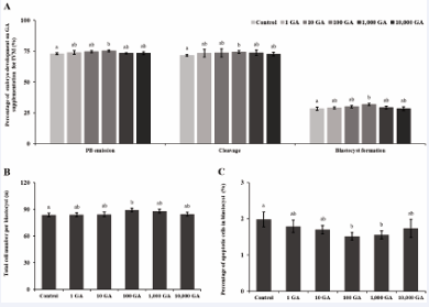
Figure 1: Effect of various concentrations of GA supplementation on the developmental capacity of parthenogenetically activated (PA) embryos. A) Polar body (PB) emission rate of MII oocytes on day 0, cleavage rate of porcine parthenogenetic embryos on day 2, and blastocyst (BL) for mation rate of PA porcine embryos on day 7. B) Total BL cell numbers on day 7. C) Percentage of BL apoptotic cells on day 7. Values are presented as means ± standard errors of the mean of inde-pendent experiments (a–bp < 0.05).
We also evaluated the PA embryo quality upon GA supplementation during porcine oocyte IVM. The total cell number per BLs was significantly higher in the 100 GA group but not in other GA groups compared with the control group (control, 83.7±2.0; 1 μM GA, 84.0±2.2; 10 μM GA, 86.1±2.1;100 μM GA, 89.3±2.1; 1 mM GA, 87.9±2.2; 10 mM GA, 84.7±2.1; p<0.05; Figure 1B). The apoptotic cell percentage was significantly higher in the control than in the 100-μM and 1-mM GA groups, but did not differ significantly in other GA groups (control, 1.98±0.2%; 1 μM GA, 1.79±0.2; 10 μM GA, 1.70±0.1; 100 μM GA, 1.51±0.1%;1 mM GA, 1.55±0.1; 10 mM GA, 1.73±0.2; p<0.05; Figure 1C). Based on these results, 100 μM GA were selected for all subsequent experiments.
Effect of GA Supplementation on the Oxidation Resistance of Porcine Oocytes Undergoing IVM
To investigate the antioxidative activity of GA supplementation during the IVM of porcine PA embryos, we measured their ROS and GSH levels and demonstrated their corresponding ROS and GSH morphologies (Figure 2A). DCFH-DA fluorescence intensity was significantly higher in the control than in the 100 GA group (control, 54.6±2.0 pixels/oocyte; 100 GA, 45.5±1.9 pixels/oocyte; p<0.01; Figure 2B). Similarly, CMF2 HC fluorescence intensity was significantly higher in the 100 GA group than in the control (control, 80.0±2.1 pixels/oocyte; 100 GA, 87.6±3.2 pixels/oocyte; p<0.05; Figure 2C). In addition, we investigated the effects of these antioxidants on the expression of the antioxidant genes, catalase (CAT), Heme Oxygenase-1 (HO-1), nuclear factor erythroid 2-like 2 (NFE2L2), superoxide dismutase 1 (SOD1), and superoxide dismutase 2 (SOD2), whose mRNA levels were normalized against those in the control group. NFE2L2, CAT, HO-1, SOD1, and SOD2 expression was significantly higher in the 100 GA group than in the control (p<0.05; Figure 2D).
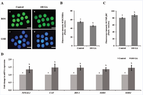
Figure 2: Effect of GA supplementation on the oxidation resistance of porcine oocytes during IVM. A) Images of oocytes stained with DCFH-DA (green) and CMF2 HC (blue). a and b, reactive oxygen species (ROS) staining; c and d, glutathione (GSH) staining; a and c, control group; b and d, 100 GA group. B) Fluorescence intensity quantification of DCFH-DA. C) Fluorescence intensity quantification of CMF2 HC. D) Relative expression of antioxidant genes (CAT, HO-1, NFE2L2, SOD1, and SOD2) at MII stage. ACTB expression served as internal control. Oocytes were analyzed at MII stage. Values are presented as means ± standard errors of the mean of independent experiments (a-bp<0.05). Scale bar, 100 μm.
Effect of GA Supplementation during IVM on Meiotic Spindle Morphology and Cytoplasmic Maturation of Porcine Oocytes
To investigate the protective effect of GA on meiotic spindle morphology of porcine oocytes, we measured chromosome alignment and spindle organization. Meiotic spindle morphology and organization have been previously studied in oocytes [27], and normal and abnormal morphologies have been characterized (Figure 3-4A). A percentage of normal chromosome alignment and meiotic spindle formation oocyte was significantly higher in the 100 GA than in the control group (control, 58.8±2.1%; GA, 68.8±2.7%; p<0.05; Figure 3B). To investigate the protective effect of GA supplementation on porcine oocyte cytoplasm maturation, we measured the p44/42 MAPK activity. Western blot analysis revealed that phospho-p44/42 MAPK, the active form of this kinase, migrated as a doublet in matured porcine oocyte lysates (Figure 3C). The ratio of phospho-p44/42 to p44/42 MAPK was normalized against that in the normal group and found to be significantly higher in the 100 GA group than in the control (control, 1.0±0.0%; GA, 1.1±0.08; p<0.05; Figure 4D).
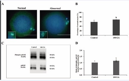
Figure 3: Effect of GA supplementation on meiotic spindle morphology and MAPK activity of porcine oocytes in vitro. A) Normal and abnormal chromosomal alignment and meiotic spindle formation in oocytes. B) Percentage of oocytes in which the chromosome morphologies and meiotic spindles were normal. C) MAPK activity determined by western blot analysis and the ratio of phospho-p44/42 to p44/42 MAPK. D) Ratio of phospho-p44/42 to p44/42 MAPK. Data were derived from 6 independent replicates per group. Data are presented as means ± standard errors of the mean (a-bp<0.05). Scale bar, 50 μm
Effect of GA Supplementation during IVM on the Quality of Porcine PA BLs
To determine whether GA supplementation during IVM affects the expression of development-related genes, we investigated the expression of developmental (caudal-related homeobox protein 2, CDX2; SRY [sex determining region Y]-box 2, SOX2; Nanog homeobox, NANOG; POU [Pit-Oct-Unc] class 5 homeobox 1; POU5F1), anti-apoptotic (Baculoviral IAP Repeat Containing 5, BIRC5; B-cell lymphoma 2 [Bcl-2]-like 1, BCL2L1), and pro apoptotic (BCL2 Antagonist/Killer 1, BAK; Fas Cell Surface Death Receptor, FAS; Caspase 3, CASP3) genes in the BL. Expression of all aforementioned developmental and anti-apoptotic genes was significantly higher in the GA 100 group than in the control (p<0.05; Figure 5). Additionally, among the pro-apoptotic genes examined, BAK, FAS, and CASP3 were significantly upregulated in the 100 GA group compared with the control (p<0.05; Figure 4).
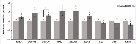
Figure 4: Effect of GA supplementation on subsequent porcine blastocyst (BL) quality during IVM. Relative expression of developmental (CDX, SOX2, NANOG, and POU5F1) and apoptotic (BIRC5, BCL2L1, BAK, FAS, and CASP3) genes in porcine embryos at BL stage. BLs were analyzed at day 7. Values are presented as means ± standard errors of the mean of independent experiments (a-bp<0.05, *p<0.01).
Effect of GA Supplementation During IVM on Porcine SCNT Embryo Developmental Competence
To investigate whether GA supplementation of porcine oocyte IVM improves the development competence of SCNTTg embryos, we assessed oocyte fusion, embryo cleavage, and BL-formation rates after SCNT. We observed that the percentage of fused oocytes was lower in control group than in the normal and 100 GA groups but did not significantly differ in other groups (normal, 67.8±1.9%; control, 66.6±2.9%; 100 GA, 67.5±1.4%; p<0.05; Figure 5A). The cleavage percentage was significantly higher in 100 GA group than in the normal and control groups (normal, 61.9±2.1%; control, 58.7±3.4%; 100 GA, 68.6±4.1%; p<0.05; Figure 5A). The BL-formation percentage was significantly higher in 100 GA group than in the normal and control groups (normal, 17.4±2.0; control, 15.1±2.6%; 100 GA, 23.8±2.1%; p<0.05; Figure 5A). In addition, we evaluated the SCNT BL quality of GA supplementation of porcine oocyte IVM. The total cell number per BL was significantly higher in the 100 GA group than in the normal and control groups (normal, 78.4±3.5; control, 73.5±3.6; 100 GA, 88.3±3.7; p<0.05; Figure 5B), and the apoptotic cell percentage was significantly higher in the control than in the GA and normal groups (normal, 0.87±0.1%; control, 1.01±0.1%; 100 GA, 0.67±0.1%; p<0.05; Figure 5C).
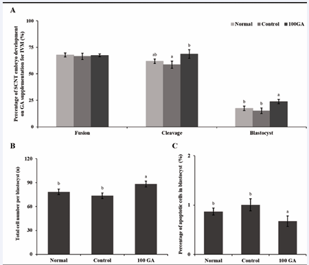
Figure 5: Effect of GA supplementation on the developmental competence of porcine SCNT embryos during IVM. A) Percentage of developed SCNT embryos upon IVM supplementation with GA. Percentages of oocytes with fusion, cleavage on day 2, and blastocyst (BL) formation on day 7 after SCNT. B) Total BL cell numbers on day 7. C) Percentage of apoptotic cells in BLs on day 7. Values are presented as means ± standard errors of the mean of independent experiments (a-bp<0.05).
DISCUSSION
In mammals, oocyte quality in optimized IVM systems has improved significantly owing to the success of ARTs, which have enhanced the development of the resulting embryos, derived from PA and SCNT cloning or Tg animals [28,29]. Therefore, recent studies have reported that supplementation of IVM with antioxidants such as melatonin, leonurine, and procyanidin B2 has efficiently reduced oocyte OS, thereby promoting its maturation and enhancing its developmental capacity [30-32]. To explore novel roles of other antioxidants on IVM, we investigated whether supplementing IVM with GA improves porcine oocyte quality and the developmental competence of the resulting embryo. We confirmed that GA acts as an antioxidant, as previously demonstrated, and by measuring a reduction in absorbance, we found that it acts by directly reducing free 2,2-diphenyl-1-picrylhydrazyl radicals. This indicates that GA can neutralize free radicals and inhibit their damaging effects. By donating an electron, GA effectively stabilizes these free radicals, thereby preventing them from causing oxidative damage to crucial cellular components such as DNA, proteins, and lipids [33 35].
This study demonstrated that PB emission rates,cleavage, and BL formation were significantly higher in the 100 GA group than in the control group (Figure 1). Previous studies have shown that GA supplementation provides effective radioprotection at 10-μM and 100-μM concentrations in lipid and aqueous phases, respectively [33]. Our results also indicated that supplementation with 100 μM GA had antioxidative effects which improved the BL-formation rate by promoting oocyte maturation. This suggests that 100 μM GA enhances the embryonic developmental competence and quality by ameliorating OS during IVM.
Several studies have indicated that early-cleavage embryos tend to develop into high-quality BLs, leading to significantly elevated pregnancy rates, implantation, and live births. Additionally, how efficiently these embryos reach the morula or BL stage correlates with early variations in the timing of their first zygotic cleavage towards the two-cell stage [36,37]. Similarly previous studies, a high percentage of cleavage correlated with high BL percentages. Therefore, 100 μM GA improved the embryonic developmental capacity and quality by reducing OS during IVM.
To assess whether GA improves embryonic development through antioxidant effects, we quantified intracellular ROS and GSH levels. The balance between intracellular oxidative and antioxidative is primarily determined by ROS and GSH activity. Controlled ROS levels promote meiotic resumption by activating the maturation factor, extruding the first polar corpuscle, and rupturing the follicular wall, thereby allowing the release of oocytes during ovulation [38,39]. Therefore, they play essential signaling roles in reproduction. Oocytes are equipped with antioxidant molecules to eliminate ROS resulting from cellular metabolism [6,40]; however, excessive ROS due to intracellular antioxidant regulatory system imbalance are a main cause of low oocyte competence [41,42]. GSH can protect oocytes from OS and is essential for cytoplasmic maturation [43]. Our data indicated that the intracellular ROS levels were significantly decreased and those of GSH were significantly increased in the 100 GA group (Figure 2B and C).
Based on this result, we demonstrated that the expression of antioxidant genes (NFE2L2, HO-1, CAT, SOD1, and SOD2) was significantly increased in the 100 GA group. The NFE2L2 transcription factor (TF) is a master regulator of cellular responses to oxidative and electrophilic stresses [44]. It controls multiple antioxidant defense mechanisms and maintains the cellular ROS balance. NFE2L2 target genes contain antioxidant response elements (AREs) that help regulate intracellular oxidant levels [45,46].
When ROS levels increase and OS occurs, antioxidant mechanisms get activated to counteract the damage. The Keap1–NFE2L2 signaling pathway is crucial for connecting cellular antioxidant response with aerobic metabolism. It regulates the cellular response to OS and redox balance by controlling NFE2L2 activation and antioxidant gene expression [47,48]. The NFE2L2/HO-1 signaling pathway activates cytoprotective genes including HO-1 against the ROS destructive effects [49]. HO-1 is a known oxidation scavenger and plays a crucial role in repairing damage caused by ROS [50]. It catalyzes the initial and rate-limiting reaction of cellular heme breakdown, thus generating an important defense product against OS [51]. Therefore, HO-1 plays a critical role in embryonic survival [52]. Free-radical scavenger enzymes such CAT and SOD have been shown to improve porcine embryo development by scavenging ROS and dampening OS [53]. SOD1 catalyzes the conversion of superoxide radicals into molecular oxygen and hydrogen peroxide [54], and is the first line of defense against the superoxide, produced as a byproduct of oxidative phosphorylation [55]. Similar to our findings, previous studies showed that GA activates NFE2L2, which triggers antioxidant enzyme synthesis. NFE2L2 binds to target-gene AREs, increasing the NFE2L2/HO-1 antioxidative pathway enzymes, CAT and SOD [56,57]. To summarize, GA improves embryonic development by inducing antioxidant effects, which are associated with decreased ROS levels and antioxidant gene expression, as well as increased GSH levels.
To assess whether GA improves embryonic development by regulating meiotic maturation, we investigated oocyte nuclear maturation. Oocyte maturation involves both nuclear and cytoplasmic maturation and is crucial for its subsequent development [58]. The regulated alignment of chromosomes and formation of a normal spindle are important for oocyte maturation [59]. OS induced the formation of an abnormal spindle, chromosome misalignment in regions where spindle-related proteins were disrupted, acetylation of α-tubulin which resulted in DNA damage, and pronounced spindle defects during the MII stage. Therefore, antioxidant supplementation could prevent the occurrence of such abnormalities during the MII stage by inhibiting OS [60,61]. We found that GA supplementation during IVM enhanced nuclear maturation by protecting the spindle and ensuring normal chromosome alignment in MII oocytes.
MAPK regulates cell cycle progression by controlling the formation of microtubules and actin filaments. It is activated upon breakdown of the germinal vesicle during oocyte maturation, and it plays a pivotal role in maintaining meiotic arrest and allowing it to resume [62].
MAPK is activated by the extracellular signal-regulated kinase (ERK) signaling pathway and participates in a crucial signal transduction cascade of extracellular signals to the nucleus [63]. Extracellular signals are transmitted to the nucleus by ERK 1/2 and induce specific transcriptional responses [64]. The phospho-p44/42 to p44/42 MAPK ratio was significantly increased in the 100 GA group, suggesting that GA supplementation during IVM improves porcine oocyte nuclear maturation.
To assess how supplementing IVM with GA affects the embryonic developmental potential, we investigated the expression of genes related to development and apoptosis. Developmental genes (CDX2, POU5F1, NANOG, and SOX2) were significantly upregulated in the 100 GA group. CDX2, a caudal-type homeodomain TF, is known to be specifically expressed in the BL trophectoderm, and its expression is sustained within the actively proliferating extra embryonic ectoderm (ExE) [65]. POU5F1 plays a crucial role to differentiate pluripotently in the inner cell mass during early embryonic development and has contributed to our understanding of how gene regulation orchestrates cellular pluripotency and facilitates progression to the BL stage [66]. NANOG, a core pioneer TF of pluripotent cells, is a strong transcriptional activator of POU5F1 expression [67,68]. SOX2 functions with POU5F1 to control the expression of target genes required for sustaining pluripotency. Its expression extends beyond pluripotent cells to regulate the ExE, trophoblast stem cells, and neural stem cells [69].
Apoptosis (programmed cell death) constitutes a specific response to OS; however, excessive apoptosis may cause early embryonic death or fetal deformities [70,71]. In the 100 GA group, anti-apoptotic genes (BCL2L and BIRC5) were significantly upregulated and pro-apoptotic genes (BAK and CASP3) were significantly downregulated, respectively. BCL2L1 supports cell survival by forming heterodimers that inhibit mitochondrial outer membrane disruption, consequently preventing the release of cytochrome c to the cytoplasm [72]. Cytochrome c, a key player in the mitochondria-dependent apoptotic pathway, activates the caspase cascade, including caspase-9 and caspase-3, which lead to apoptosis [73]. BIRC5 has been shown to downregulate programmed cell death by inhibiting caspase activation [74]. Also, BCL2L1 and BIRC5 can inhibit apoptosis in mitochondria [75]. By contrast, BAK triggers mitochondrial outer membrane permeabilization, leading to the release of cytochrome c into the cytoplasm and subsequent activation of the apoptotic pathway [76]. CASP3 facilitates the degradation of essential matrix proteins and initiates morphological and biochemical alterations that are characteristic of apoptosis.
These changes include DNA fragmentation and chromosome condensation [76,77]. This suggests that supplementation of IVM with GA may exert positive effects on maintaining embryonic pluripotency, normal embryonic development, and the inhibition of apoptosis.
Based on previous experiments showing that GA enhances oocyte maturation and subsequent embryonic developmental rates by means of its antioxidant activity, we investigated whether it also enhances subsequent SCNT embryonic development using normal and transduction donor cells. The developmental capacity of SCNT embryos using Tg cells was similar to that using non-Tg cells [78]. Also, SCNT embryos showed low developmental competence compared with PA embryos [79,80]. This may be related to the SCNT technique, genetic background, or incorrect epigenetic reprogramming of the donor cell nuclei [81,82]. Nonetheless, the 100 GA supplementation enhanced the development and BL quality of SCNT embryos compared with the normal and control groups. Improving the recipient oocyte quality is important for efficient epigenetic reprogramming of donor cell nuclei [83]. Previous studies have demonstrated that the recipient cytoplasm influences both BL formation and fetal survival l [84]. These results suggest that the GA supplementation improves the developmental capacity and quality of porcine embryos by protecting OS during IVM.
FUNDING
This study demonstrated that supplementation with 100 μM GA enhanced the oocyte maturation and subsequent developmental capacity of porcine PA and SCNT embryos. These findings suggest that GA supplementation may help improve IVM in ART and biotechnology, as well as increase its potential for clinical
This work was supported by a grant (RS-2023 00254212) from the Technology Commercialization Support Program, Ministry of Agriculture, Food and Rural Affairs, Republic of Korea.
REFERENCES
- Wilmut I, Schnieke AE, McWhir J, Kind AJ, Campbell KH. Viable offspring derived from fetal and adult mammalian cells. Nature. 1997; 385: 810-813.
- Han K, Liang L, Li L, Ouyang Z, Zhao B, Wang Q, et al. Generation of Hoxc13 knockout pigs recapitulates human ectodermal dysplasia-9. Hum Mol Genet. 2017; 26: 184-191.
- Davis BT, Wang XJ, Rohret JA, Struzynski JT, Merricks EP, Bellinger DA, et al. Targeted disruption of LDLR causes hypercholesterolemia and atherosclerosis in Yucatan miniature pigs. PLoS One. 2014; 9: e93457.
- Lunney JK, Van Goor A, Walker KE, Hailstock T, Franklin J, Dai C. Importance of the pig as a human biomedical model. Sci Transl Med. 2021; 13: eabd5758.
- Lee SE, Hyun H, Park MR, Choi Y, Son YJ, Park YG, et al. Production of transgenic pig as an Alzheimer’s disease model using a multi-cistronic vector system. PLoS One. 2017; 12: e0177933.
- Kala M, Shaikh MV, Nivsarkar M. Equilibrium between anti-oxidants and reactive oxygen species: a requisite for oocyte development and maturation. Reprod Med Biol. 2017; 16: 28-35.
- Schmidt M, Winther KD, Secher JO, Callesen H. Postmortem findings in cloned and transgenic piglets dead before weaning. Theriogenology. 2015; 84: 1014-1023.
- Manjunatha BM, Gupta PS, Devaraj M, Ravindra JP, Nandi S. Selection of developmentally competent buffalo oocytes by brilliant cresyl blue staining before IVM. Theriogenology. 2007; 68: 1299-1304.
- ?Kurome M, Geistlinger L, Kessler B, Zakhartchenko V, Klymiuk N, Wuensch A, et al. Factors influencing the efficiency of generating genetically engineered pigs by nuclear transfer: multi-factorial analysis of a large data set. BMC Biotechnol. 2013; 13: 43.
- Marchal R, Tomanek M, Terqui M, Mermillod P. Effects of cell cycle dependent kinases inhibitor on nuclear and cytoplasmic maturation of porcine oocytes. Mol Reprod Dev. 2001; 60: 65-73.
- Yan K, Cui K, Nie J, Zhang H, Sui L, Zhang H, et al. Mogroside V Protects Porcine Oocytes From Lipopolysaccharide-Induced Meiotic Defects. Front Cell Dev Biol. 2021; 9: 639691.
- Lee Y, Lee J, Hyun SH, Lee GS, Lee E. In vitro maturation using αMEM with reduced NaCl enhances maturation and developmental competence of pig oocytes after somatic cell nuclear transfer. J Vet Sci. 2022; 23: e31.
- Son J, Malaweera DB, Lee E, Shin S, Cho J. Development of in vitro produced porcine embryos according to serum types as macromolecule. J Vet Sci. 2013; 14: 315-321.
- Nie J, Yan K, Sui L, Zhang H, Zhang H, Yang X, et al. Mogroside V improves porcine oocyte in vitro maturation and subsequent embryonic development. Theriogenology. 2020; 141: 35-40.
- Lee SH. Human Adipose-Derived Stem Cells’ Paracrine Factors in Conditioned Medium Can Enhance Porcine Oocyte Maturation and Subsequent Embryo Development. Int J Mol Sci. 2021; 22: 579.
- Nadeem MS, Kazmi I, Ullah I, Muhammad K, Anwar F. Allicin, an Antioxidant and Neuroprotective Agent, Ameliorates Cognitive Impairment. Antioxidants (Basel). 2021; 11: 87.
- Godoy-Caballero MP, Acedo-Valenzuela MI, Galeano-Díaz T. Simple quantification of phenolic compounds present in the minor fraction of virgin olive oil by LC-DAD-FLD. Talanta. 2012; 101: 479-487.
- Harris CS, Mo F, Migahed L, Chepelev L, Haddad PS, Wright JS, et al. Plant phenolics regulate neoplastic cell growth and survival: a quantitative structure-activity and biochemical analysis. Can J Physiol Pharmacol. 2007; 85: 1124-1138.
- Sezer ED, Oktay LM, Karadada? E, Memmedov H, Selvi Gunel N, Sözmen E. Assessing Anticancer Potential of Blueberry Flavonoids, Quercetin, Kaempferol, and Gentisic Acid, Through Oxidative Stress and Apoptosis Parameters on HCT-116 Cells. J Med Food. 2019; 22: 1118-1126.
- Abedi F, Razavi BM, Hosseinzadeh H. A review on gentisic acid as a plant derived phenolic acid and metabolite of aspirin: Comprehensive pharmacology, toxicology, and some pharmaceutical aspects. Phytother Res. 2020; 34: 729-741.
- Noei Razliqi R, Ahangarpour A, Mard SA, Khorsandi L. Gentisic acid protects against diabetic nephropathy in Nicotinamide-Streptozotocin administered male mice by attenuating oxidative stress and inflammation: The role of miR-200a/Keap1/Nrf2 pathway, renin-angiotensin system (RAS) and NF-?B. Chem Biol Interact. 2023; 380: 110507.
- Hinz B, Kraus V, Pahl A, Brune K. Salicylate metabolites inhibit cyclooxygenase-2-dependent prostaglandin E(2) synthesis in murine macrophages. Biochem Biophys Res Commun. 2000; 274: 197-202.
- Ashidate K, Kawamura M, Mimura D, Tohda H, Miyazaki S, Teramoto T, et al. Gentisic acid, an aspirin metabolite, inhibits oxidation of low-density lipoprotein and the formation of cholesterol ester hydroperoxides in human plasma. Eur J Pharmacol. 2005; 513: 173-179.
- Kim M, Kim J, Shin YK, Kim KY. Gentisic Acid Stimulates Keratinocyte Proliferation through ERK1/2 Phosphorylation. Int J Med Sci. 2020; 17: 626-631.
- Lee SE, Sun SC, Choi HY, Uhm SJ, Kim NH. mTOR is required for asymmetric division through small GTPases in mouse oocytes. Mol Reprod Dev. 2012; 79: 356-366.
- Livak KJ, Schmittgen TD. Analysis of relative gene expression data using real-time quantitative PCR and the 2(-Delta Delta C(T)) Method. Methods. 2001; 25: 402-408.
- Lenie S, Cortvrindt R, Eichenlaub-Ritter U, Smitz J. Continuous exposure to bisphenol A during in vitro follicular development induces meiotic abnormalities. Mutat Res. 2008; 651: 71-81.
- Wang Y, Qi JJ, Yin YJ, Jiang H, Zhang JB, Liang S, et al. Ferulic Acid Enhances Oocyte Maturation and the Subsequent Development of Bovine Oocytes. Int J Mol Sci. 2023; 24: 14804.
- Keim J, Liu Y, Regouski M, Stott R, Singina GN, White KL, et al. Cytokine supplemented maturation medium improved development to term following somatic cell nuclear transfer (SCNT) in cattle. Reprod Fertil Dev. 2023; 35: 575-588.
- Yang D, Mu Y, Wang J, Zou W, Zou H, Yang H, Zhang C, Fan Y, Zhang H, Zhang H, Chen B, Zhang Z. Melatonin enhances the developmental potential of immature oocytes from older reproductive-aged women by improving mitochondrial function. Heliyon. 2023; 9: e19366.
- Zheng L, Luo Y, Zhou D, Liu H, Zhou G, Meng L, et al. Leonurine improves bovine oocyte maturation and subsequent embryonic development by reducing oxidative stress and improving mitochondrial function. Theriogenology. 2023; 199: 11-18.
- Bai J , Li J , Wang L , Hao S , Guo Y , Liu Y , et al. Effect of antioxidant procyanidin b2 (pcb2) on ovine oocyte developmental potential in response to in vitro maturation (ivm) and vitrification stress. Cryo Letters. 2023; 44: 109-117.
- Joshi R, Gangabhagirathi R, Venu S, Adhikari S, Mukherjee T. Antioxidant activity and free radical scavenging reactions of gentisic acid: in-vitro and pulse radiolysis studies. Free Radic Res. 2012; 46: 11-20.
- Mardani-Ghahfarokhi A, Farhoosh R. Antioxidant activity and mechanism of inhibitory action of gentisic and α-resorcylic acids. Sci Rep. 2020; 10: 19487.
- Villaño D, Fernández-Pachón MS, Moyá ML, Troncoso AM, García-Parrilla MC. Radical scavenging ability of polyphenolic compounds towards DPPH free radical. Talanta. 2007; 71: 230-235.
- Isom SC, Li RF, Whitworth KM, Prather RS. Timing of first embryonic cleavage is a positive indicator of the in vitro developmental potential of porcine embryos derived from in vitro fertilization, somatic cell nuclear transfer and parthenogenesis. Mol Reprod Dev. 2012; 79: 197-207.
- Yaacobi-Artzi S, Kalo D, Roth Z. Association between the morphokinetics of in-vitro-derived bovine embryos and the
transcriptomic profile of the derived blastocysts. PLoS One. 2022; 17: e0276642.
- Pandey AN, Chaube SK. A moderate increase of hydrogen peroxide level is beneficial for spontaneous resumption of meiosis from diplotene arrest in rat oocytes cultured in vitro. Biores Open Access. 2014; 3: 183-191.
- Tiwari M, Chaube SK. Moderate increase of reactive oxygen species triggers meiotic resumption in rat follicular oocytes. J Obstet Gynaecol Res. 2016; 42: 536-546.
- Mihalas BP, Redgrove KA, McLaughlin EA, Nixon B. Molecular Mechanisms Responsible for Increased Vulnerability of the Ageing Oocyte to Oxidative Damage. Oxid Med Cell Longev. 2017; 2017: 4015874.
- Mahrous E, Yang Q, Clarke HJ. Regulation of mitochondrial DNA accumulation during oocyte growth and meiotic maturation in the mouse. Reproduction. 2012; 144: 177-185.
- Wang S, He G, Chen M, Zuo T, Xu W, Liu X. The Role of Antioxidant Enzymes in the Ovaries. Oxid Med Cell Longev. 2017; 2017: 4371714.
- Luberda Z. The role of glutathione in mammalian gametes. Reprod Biol. 2005; 5: 5-17.
- Baird L, Yamamoto M. The Molecular Mechanisms Regulating the KEAP1-NRF2 Pathway. Mol Cell Biol. 2020 Jun 15;40(13):e00099-e00120.
- Motohashi H, Yamamoto M. Nrf2–Keap1 defines a physiologically important stress response mechanism. Trends Mol Med. 2004; 10: 549-57.
- Itoh K, Ishii T, Wakabayashi N, Yamamoto M. Regulatory mechanisms of cellular response to oxidative stress. Free Radical Res. 1999; 31: 319-324.
- Liang Y, Batistel F, Parys C, Loor J. Glutathione metabolism and nuclear factor erythroid 2-like 2 (NFE2L2)-related proteins in adipose tissue are altered by supply of ethyl-cellulose rumen-protected methionine in peripartal Holstein cows. J Dairy Sci. 2019; 102: 5530-5541.
- Kensler TW, Wakabayashi N, Biswal S. Cell survival responses to environmental stresses via the Keap1-Nrf2-ARE pathway. Annu Rev Pharmacol Toxicol. 2007; 47: 89-116.
- Fan X, Wei W, Huang J, Peng L, Ci X. Daphnetin attenuated cisplatin-induced acute nephrotoxicity with enhancing antitumor activity of cisplatin by upregulating SIRT1/SIRT6-Nrf2 pathway. Front Pharmacol. 2020; 11: 579178.
- Abdel-Wahab BA, Walbi IA, Albarqi HA, Ali FE, Hassanein EH. Roflumilast protects from cisplatin-induced testicular toxicity in male rats and enhances its cytotoxicity in prostate cancer cell line. Role of NF-κB-p65, cAMP/PKA and Nrf2/HO-1, NQO1 signaling. Food and Chemical Toxicol. 2021; 151: 112133.
- Drummond GS, Baum J, Greenberg M, Lewis D, Abraham NG. HO-1 overexpression and underexpression: Clinical implications. Arch Biochem Biophys. 2019; 673: 108073.
- Lai YL, Lin CY, Jiang WC, Ho YC, Chen CH, Yet SF. Loss of heme oxygenase-1 accelerates mesodermal gene expressions during embryoid body development from mouse embryonic stem cells. Redox Biology. 2018; 15: 51-61.
- Takahashi M. Oxidative stress and redox regulation on in vitro development of mammalian embryos. J Reproduction and Development. 2012; 58: 1-9.
- McCord JM, Fridovich I. Superoxide dismutase: an enzymic function for erythrocuprein (hemocuprein). J Biol Chem. 1969; 244: 6049-6055.
- Li Y, Huang T-T, Carlson EJ, Melov S, Ursell PC, Olson JL, et al. Dilated cardiomyopathy and neonatal lethality in mutant mice lacking manganese superoxide dismutase. Nature genetics. 1995; 11: 376-381.
- Altinoz MA, Elmaci I, Cengiz S, Emekli-Alturfan E, Ozpinar A. From epidemiology to treatment: Aspirin’s prevention of brain and breast-cancer and cardioprotection may associate with its metabolite gentisic acid. Chemico-biological interactions. 2018; 291: 29-39.
- Dachineni R, Kumar DR, Callegari E, Kesharwani SS, Sankaranarayanan R, Seefeldt T, et al. Salicylic acid metabolites and derivatives inhibit CDK activity: Novel insights into aspirin’s chemopreventive effects against colorectal cancer. Int J Oncol. 2017; 51: 1661-1673.
- Akar Y, Ahmad N, Khal?d M. The effect of cadmium on the bovine in vitro oocyte maturation and early embryo development. Int J Vet Sci Med. 2018; 6: S73-S7.
- Clift D, Schuh M. A three-step MTOC fragmentation mechanism facilitates bipolar spindle assembly in mouse oocytes. Nat Commun. 2015; 6: 7217.
- Han L, Wang H, Li L, Li X, Ge J, Reiter RJ, et al. Melatonin protects against maternal obesity-associated oxidative stress and meiotic defects in oocytes via the SIRT 3-SOD 2-dependent pathway. J Pineal Res. 2017; 63: e12431.
- Wang G-F, Dong Q, Bai Y, Yuan J, Xu Q, Cao C, et al. Oxidative stress induces mitotic arrest by inhibiting Aurora A-involved mitotic spindle formation. Free Radic Biol Med. 2017; 103: 177-187.
- Villa-Diaz LG, Miyano T. Activation of p38 MAPK during porcine oocyte maturation. Biol Reproduction. 2004; 71: 691-696.
- Michael J, Wurtzel J, Goldfinger L. Regulation of H-Ras-driven MAPK signaling, transformation and tumorigenesis, but not PI3K signaling and tumor progression, by plasma membrane microdomains. Oncogenesis. 2016; 5: e228.
- Robinson MJ, Stippec SA, Goldsmith E, White MA, Cobb MH. A constitutively active and nuclear form of the MAP kinase ERK2 is sufficient for neurite outgrowth and cell transformation. Curr Biol. 1998; 8: 1141-1152.
- Strumpf D, Mao CA, Yamanaka Y, Ralston A, Chawengsaksophak K, Beck F,et al. Cdx2 is required for correct cell fate specification and differentiation of trophectoderm in the mouse blastocyst. Development. 2005; 132: 2093-2102.
- Nichols J, Zevnik B, Anastassiadis K, Niwa H, Klewe-Nebenius D, Chambers I, et al. Formation of pluripotent stem cells in the mammalian embryo depends on the POU transcription factor Oct4. Cell. 1998; 95: 379-391.
- Pan G, Li J, Zhou Y, Zheng H, Pei D, Pan G, et al. A negative feedback loop of transcription factors that controls stem cell pluripotency and self-renewal. The FASEB J. 2006; 20: 1730-1732.
- Plachta N, Bollenbach T, Pease S, Fraser SE, Pantazis P. Oct4 kinetics predict cell lineage patterning in the early mammalian embryo. Nature Cell Biology. 2011; 13: 117-123.
- Avilion AA, Nicolis SK, Pevny LH, Perez L, Vivian N, Lovell-Badge R. Multipotent cell lineages in early mouse development depend on SOX2 function. Genes Dev. 2003; 17: 126-140.
- Parrish AB, Freel CD, Kornbluth S. Cellular mechanisms controlling caspase activation and function. Cold Spring Harb Perspect Biol . 2013; 5: a008672.
- Brison DR, Schultz RM. Apoptosis during mouse blastocyst formation: evidence for a role for survival factors including transforming growth factor α. Biol Reprod. 1997; 56: 1088-1096.
- Scott A, Khan K, Heer J, Cook J, Lian O, Duronio V. High strain mechanical loading rapidly induces tendon apoptosis: an ex vivo rat tibialis anterior model. Br J Sports Med. 2005; 39: e25.
- White E. Life, death, and the pursuit of apoptosis. Genes dev. 1996;10: 1-15.
- Sun X, Zhang H, Zhang Y, Yang Q, Zhao S. Caspase-dependent mitochondrial apoptotic pathway is involved in astilbin-mediated cytotoxicity in breast carcinoma cells. Oncology Rep. 2018; 40: 2278-2286.
- De Felici M, Di Carlo A, Pesce M, Iona S, Farrace M, Piacentini M. Bcl-2 and Bax regulation of apoptosis in germ cells during prenatal oogenesis in the mouse embryo. Cell Death Differ. 1999; 6: 908-915.
- Reed JC. Double identity for proteins of the Bcl-2 family. Nature. 1997; 387: 773-776.
- Renault TT, Floros KV, Chipuk JE. BAK/BAX activation and cytochrome c release assays using isolated mitochondria. Methods. 2013; 61: 146-155.
- sang Lee G, soo Kim H, hwan Hyun S, hyun Lee S, yong Jeon H, hyun Nam D, et al. Production of transgenic cloned piglets from genetically transformed fetal fibroblasts selected by green fluorescent protein. Theriogenology. 2005; 63: 973-991.
- Lee H-B, Lee S-E, Park M-J, Han D-H, Lim E-S, Ryu B, et al. Ellagic acid treatment during in vitro maturation of porcine oocytes improves development competence after parthenogenetic activation and somatic cell nuclear transfer. Theriogenology. 2024; 215: 214-223
- Pyeon D-B, Lee S-E, Yoon J-W, Park H-J, Oh S-H, Lee D-G, et al. Comparison of the improving embryo development effects of Sasa quelpaertensis Nakai extract, p-coumaric acid, and myricetin on porcine oocytes according to their antioxidant capacities. Theriogenology. 2022; 185: 97-108.
- Diao YF, Oqani RK, Li XX, Lin T, Kang JW, Jin DI. Changes in histone H3 lysine 36 methylation in porcine oocytes and preimplantation embryos. PloS one. 2014; 9: e100205.
- Oh S-H, Lee S-E, Han D-H, Yoon J-W, Kim S-H, Lim E-S, et al. Treatments of Porcine Nuclear Recipient Oocytes and Somatic Cell Nuclear Transfer-Generated Embryos with Various Reactive Oxygen Species Scavengers Lead to Improvements of Their Quality Parameters and Developmental Competences by Mitigating Oxidative Stress-Related Impacts. Cellular Reprogramming. 2023; 25: 73-81.
- Miyoshi K, Rzucidlo SJ, Pratt SL, Stice SL. Improvements in cloning efficiencies may be possible by increasing uniformity in recipient oocytes and donor cells. Biol. Reprod. 2003; 68: 1079-1086.
- Naoi H, Agung B, Karja N, Wongsrikeao P, Shimizu R, Taniguchi M, et al. Effects of the reproductive status on morphological oocyte quality and developmental competence of oocytes after in vitro fertilization and somatic cell nuclear transfer in cat. Reprod Domest Anim. 2008; 43: 157-161.applications.